Copyright
©2008 The WJG Press and Baishideng.
World J Gastroenterol. Jul 28, 2008; 14(28): 4473-4479
Published online Jul 28, 2008. doi: 10.3748/wjg.14.4473
Published online Jul 28, 2008. doi: 10.3748/wjg.14.4473
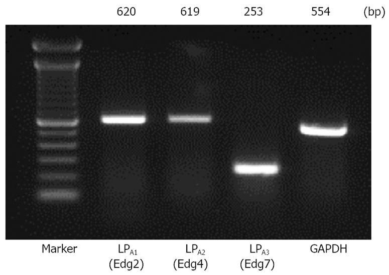
Figure 1 Expression of LPA receptors in Panc-1 cells.
LPA-specific receptors LPA1/Edg-2, LPA2/Edg-4, and LPA3/Edg-7 are expressed in PANC-1 cells. Total mRNA of PANC-1 cells was extracted and used for RT and PCR using primers designed from the sequence of LPA1, LPA2, and LPA3, and GAPDH. PCR products were separated by electrophoresis in 2.0% agarose gel. The molecular size of the amplification products is inferred from the electrophoretic migration of the DNA markers.
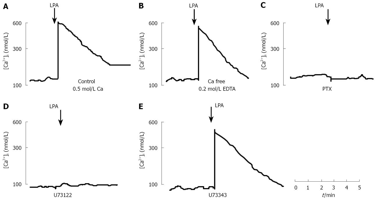
Figure 2 Effect of LPA on cytosolic free calcium concentration [Ca2+]i in Panc-1 cells.
A: Representative Ca2+ signal was evoked by 10 &mgr;mol/L LPA in Panc-1 cells; B: Effect of chelation of extracellular Ca2+ by addition of 0.2 mmol/L EDTA on LPA-induced increases in Ca2+; C: PTX-sensitive effect of LPA on cytosolic free calcium. Panc-1 cells were pretreated with 100 ng/mL of PTX for overnight and loaded with fura-2/AM; D: Effect of U73122, a phospholipase C inhibitor, on LPA-induced increases in cytosolic free calcium in Panc-1 cells. Panc-1 cells were treated with U73122 at a concentration of 10 &mgr;mol/L for 10 min, and then stimulated by 10 &mgr;mol/L LPA; E: Effect of U73343, an inactive analogue of U73122, on LPA-evoked increases in cytosolic free calcium in Panc-1 cells.
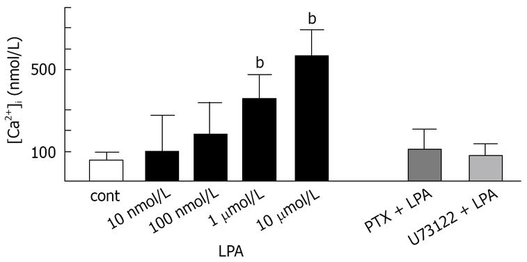
Figure 3 Effect of LPA on cytosolic free calcium in Panc-1.
LPA evoked cytosolic free calcium in concentration-dependent manner. PTX-treatment at a concentration of 100 ng/ml for overnight abolished 10 &mgr;mol/L LPA-induced mobilization of [Ca2+]i. Pretreatment of U73122 (10 &mgr;mol/L) for 10 min also abolished LPA-induced mobilization of [Ca2+]i. bP < 0.01.
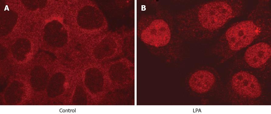
Figure 4 Effect of LPA on nuclear translocation of NF-κB.
LPA-induced nuclear translocation of p65 in Panc-1 cells. Localization of p65 was visualized by indirect immunofluorescence staining using rabbit anti-p65 polyclonal antibodies (1:50) which only recognized NF-κB p65. Donkey anti-rabbit antibodies (1:100) conjugated to rhodamine was performed, and visualized under a confocal microscope. A: Cytosolic localization of p65 in an inactive form in unstimulated Panc-1 cells; B: Nuclear localization of p65 in Panc-1 cells after treatment of 10 &mgr;mol/L LPA was observed at 60 min.
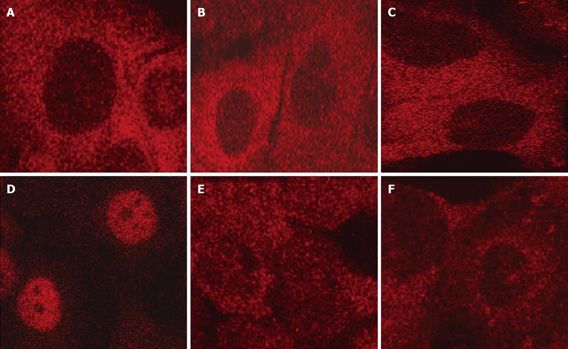
Figure 5 Localization of NF-κB p65 in Panc-1 cells.
A: PTX-treatment prevented nuclear translocation of NF-κB p65 induced by LPA in Panc-1 cells at 1 h. Panc-1 cells were preincubated with 100 ng/mL PTX, and then stimulated with 10 &mgr;mol/L LPA; B: U73122-treatment prevented nuclear translocation of NF-κB p65 induced by LPA in Panc-1 cells at 1 h. Panc-1 cells were pretreated with 10 &mgr;mol/L U73122 for 10 min before adding 10 &mgr;mol/L LPA; C: Nuclear translocation of p65 induced by LPA was reduced by an intracellular calcium chelator, BPTA-AM at 1 h. Panc-1 cells were treated with 10 &mgr;mol/L BAPTA-AM for 15 min and then stimulated with 10 &mgr;mol/L LPA; D: Thapsigargin alone caused nuclear translocation of p65 at 1 h. Panc-1 cells were treated with 500 nmol/L thapsigarigin; E: Phorbol myristate (PMA) alone failed to mobilize p65 into nuclei at 1 h. Panc-1 cells were treated with 100 nmol/L PMA; F: Staurosporine reduced nuclear localization of p65 induced by LPA at 1 h. Panc-1 cells were treated with 10 nmol/L staurosporine for 10 min and then stimulated with 10 &mgr;mol/L LPA.
- Citation: Arita Y, Ito T, Oono T, Kawabe K, Hisano T, Takayanagi R. Lysophosphatidic acid induced nuclear translocation of nuclear factor-κB in Panc-1 cells by mobilizing cytosolic free calcium. World J Gastroenterol 2008; 14(28): 4473-4479
- URL: https://www.wjgnet.com/1007-9327/full/v14/i28/4473.htm
- DOI: https://dx.doi.org/10.3748/wjg.14.4473


