Copyright
©2008 The WJG Press and Baishideng.
World J Gastroenterol. Jul 21, 2008; 14(27): 4319-4323
Published online Jul 21, 2008. doi: 10.3748/wjg.14.4319
Published online Jul 21, 2008. doi: 10.3748/wjg.14.4319
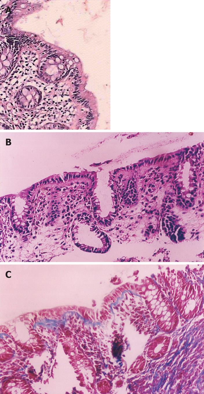
Figure 1 Pathologic view (× 200).
A: Lymphocytic colitis. Note the increased number of chronic inflammatory cells in the lamina propria and within the surface epithelium (HE); B: Collagenous colitis. Note the subepithelial thick collagenous band (HE); C: Collagenous band thickness on Mason trichrome dye.
- Citation: Erdem L, Yildirim S, Akbayir N, Yilmaz B, Yenice N, Gültekin OS, Peker &. Prevalence of microscopic colitis in patients with diarrhea of unknown etiology in Turkey. World J Gastroenterol 2008; 14(27): 4319-4323
- URL: https://www.wjgnet.com/1007-9327/full/v14/i27/4319.htm
- DOI: https://dx.doi.org/10.3748/wjg.14.4319


