Copyright
©2008 The WJG Press and Baishideng.
World J Gastroenterol. Jul 14, 2008; 14(26): 4253-4256
Published online Jul 14, 2008. doi: 10.3748/wjg.14.4253
Published online Jul 14, 2008. doi: 10.3748/wjg.14.4253
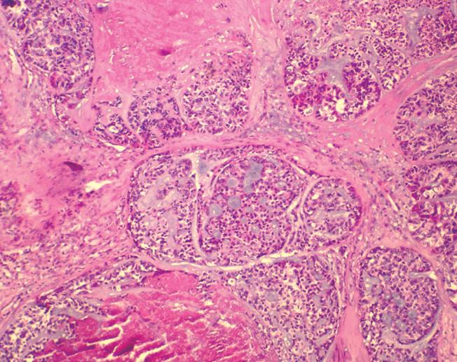
Figure 1 Arrangement of basaloid cells in the form of anastomosing trabeculae and microcystic structures.
The microcystic spaces contained basophilic mucoid matrix, mimicking an adenoid cystic carcinoma. The cells at the edges of basaloid islands tended to show peripheral nuclear palisading. Comedo necrosis was observed in the center of basaloid lobules (HE, × 40).
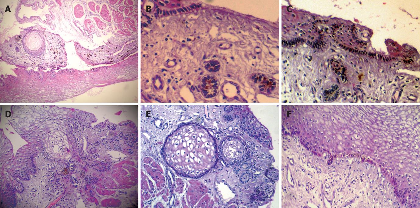
Figure 2 Focal distribution of melanocytes in keratinocytes of the basal esophageal mucosa layer, heavily pigmented, spindled or dendritic melanocytes/pigmentophages locate predominately in the superficial lamina propria, Moreover, there are several hair follicles and a horn cyst in the mucosa, with melanocytes in the hair follicle (HE, × 40) (A), and distribution of melanocytes in keratinocytes of the basal esophageal mucosa layer and hair follicles in the mucosal lamina propria with melanocytes (B,C) (B: HE, × 200; C: HE, × 40), cluster of hair follicles and horn cysts in the mucosa (D,E) (HE, × 40), and focal distribution of melanocytes in keratinocytes of the basal esophageal mucosa layer below the BSC (HE, × 40) (F).
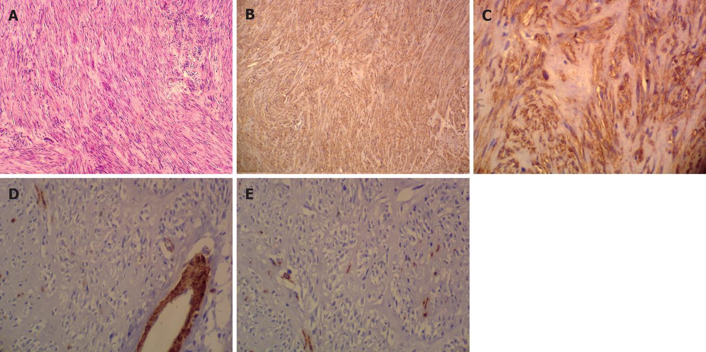
Figure 3 Gastric tumor consisting of basophilic spindle-cells arranged in short fascicles and aligned in a strikingly Schwannian pattern with prominent nuclear palisading but no mitotic activity and necrosis (HE, × 40) (A), and immunohistochemistry showing strong immunoreactivity of basophilic spindle-cells on CD117 (B) (× 40) and CD34 (C) (× 400) but no specific reaction to the antibodies SMA (D) (× 200) and S-100 (E) (× 200).
- Citation: Wang DG, Li XG, Gao H, Sun XY, Zhou XQ. Coexistence of esophageal blue nevus, hair follicles and basaloid squamous carcinoma: A case report. World J Gastroenterol 2008; 14(26): 4253-4256
- URL: https://www.wjgnet.com/1007-9327/full/v14/i26/4253.htm
- DOI: https://dx.doi.org/10.3748/wjg.14.4253


