Copyright
©2008 The WJG Press and Baishideng.
World J Gastroenterol. Jul 14, 2008; 14(26): 4249-4252
Published online Jul 14, 2008. doi: 10.3748/wjg.14.4249
Published online Jul 14, 2008. doi: 10.3748/wjg.14.4249
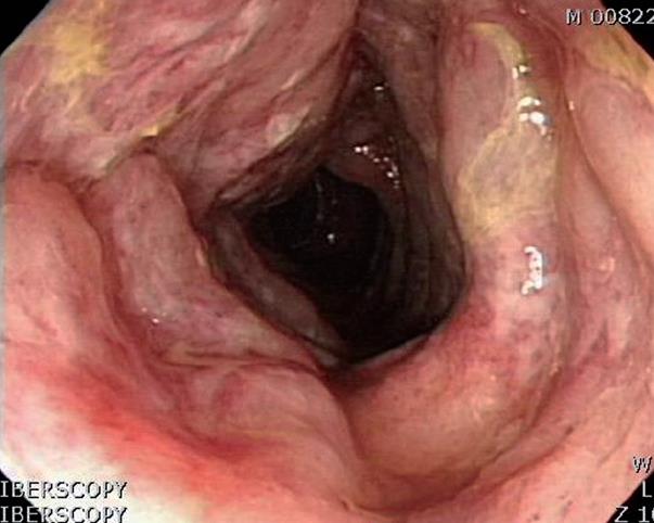
Figure 1 Colonoscopy revealing diffuse colonic ulceration, exudate, and hemorrhage in the sigmoid and descending colon.
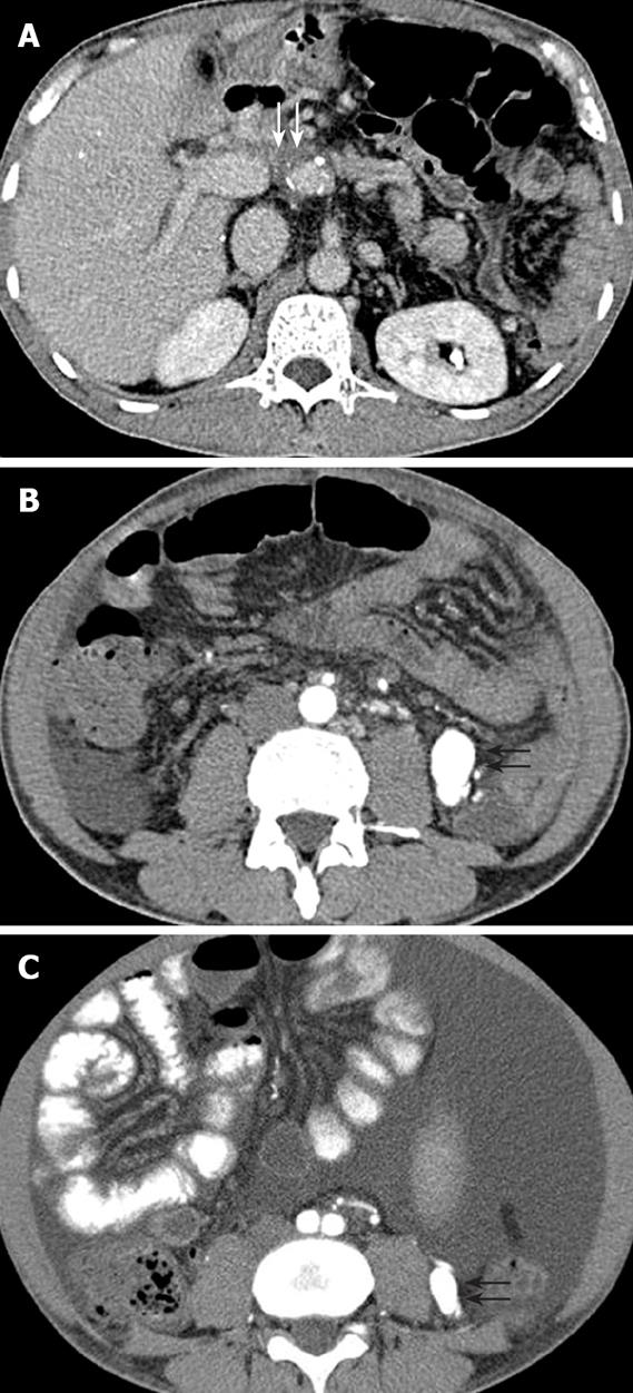
Figure 2 Contrast-enhanced CT of the abdomen showing portal vein stenosis (white arrows) (A), an approximately 30 mm × 18 mm contrast-enhancing vascular mass (black arrows) (B), and an approximately 10 mm × 8 mm enhancing vascular structure before transplantation (black arrows) (C).
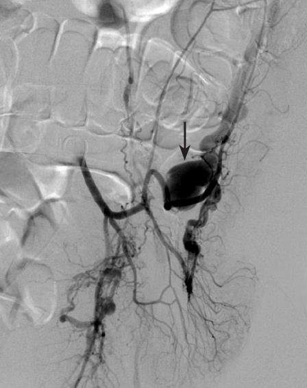
Figure 3 Angiography of the inferior mesenteric artery showing an AVF (arrow) with early opacification of a dilated vein.
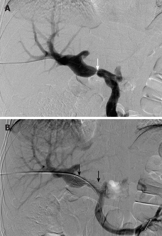
Figure 4 Transhepatic portal venography showing portal vein stenosis (white arrow) with stasis in the mesenteric vein (A) and good patency of the portal vein following deployment of the metal stent (black arrows) (B).
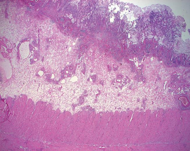
Figure 5 Histopathologic examination of the resected sigmoid showing diffuse ischemic necrosis inflammatory infiltrations (HE × 20).
- Citation: Kim IH, Kim DG, Kwak HS, Yu HC, Cho BH, Park HS. Ischemic colitis secondary to inferior mesenteric arteriovenous fistula and portal vein stenosis in a liver transplant recipient. World J Gastroenterol 2008; 14(26): 4249-4252
- URL: https://www.wjgnet.com/1007-9327/full/v14/i26/4249.htm
- DOI: https://dx.doi.org/10.3748/wjg.14.4249


