Copyright
©2008 The WJG Press and Baishideng.
World J Gastroenterol. Jul 7, 2008; 14(25): 4070-4076
Published online Jul 7, 2008. doi: 10.3748/wjg.14.4070
Published online Jul 7, 2008. doi: 10.3748/wjg.14.4070
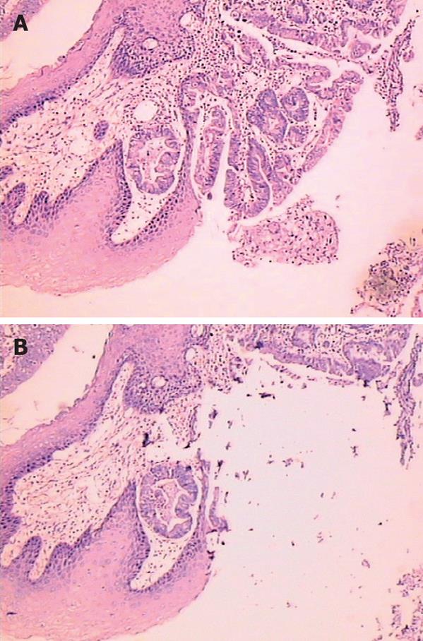
Figure 1 Photomicrograph of epithelial dysplasia (HE, × 200) in Barrett esophagus (A) and after dissected under microscopic guidance (B).
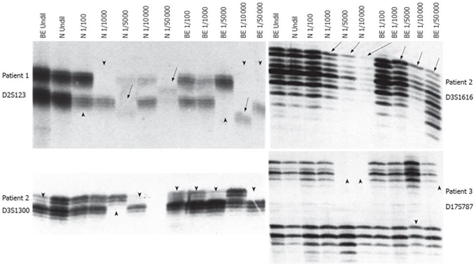
Figure 2 Microsatellite alterations in diluted DNA.
Arrow and arrowhead indicate MSI and LOH, respectively. N: Normal esophageal squamous epithelium DNA; BE: Barrett esophageal columnar epithelium (metaplasia) DNA; Undil: Un-diluted; 1/100-1/50 000: Dilution fold.
- Citation: Cai JC, Liu D, Liu KH, Zhang HP, Zhong S, Xia NS. Microsatellite alterations in phenotypically normal esophageal squamous epithelium and metaplasia-dysplasia-adenocarcinoma sequence. World J Gastroenterol 2008; 14(25): 4070-4076
- URL: https://www.wjgnet.com/1007-9327/full/v14/i25/4070.htm
- DOI: https://dx.doi.org/10.3748/wjg.14.4070


