Copyright
©2008 The WJG Press and Baishideng.
World J Gastroenterol. Jul 7, 2008; 14(25): 3982-3989
Published online Jul 7, 2008. doi: 10.3748/wjg.14.3982
Published online Jul 7, 2008. doi: 10.3748/wjg.14.3982
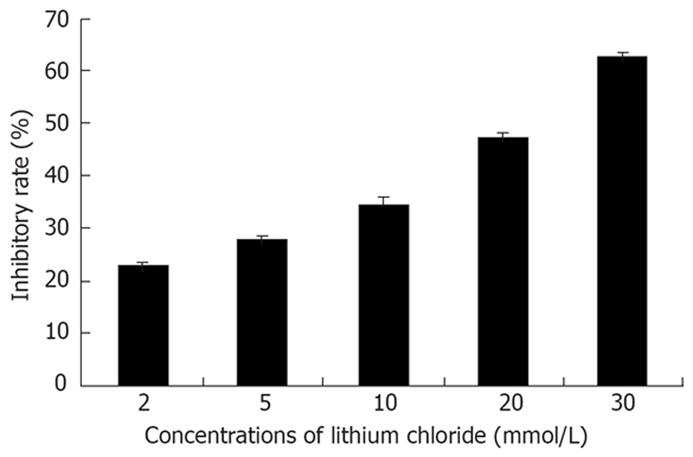
Figure 1 Lithium could impose inhibition on Eca-109 cells in day 3.
With the increased concentration of lithium, the inhibitory effect was enhanced. Each bar came from six wells of 96-well plate and represented mean ± SD.
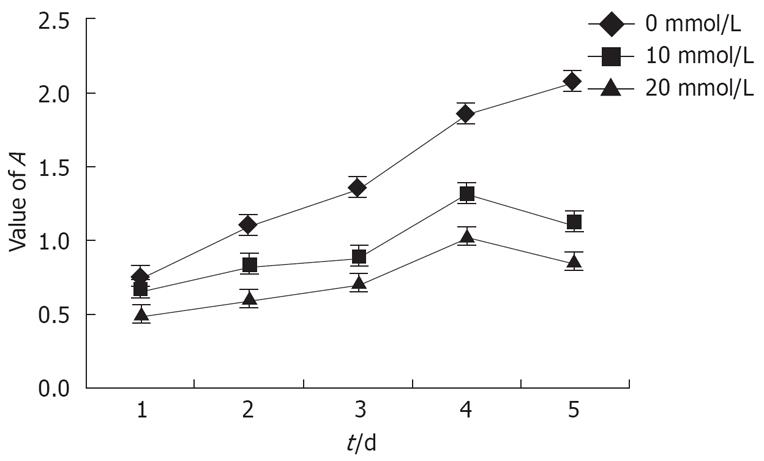
Figure 2 Growth curves of Eca-109 cells plotted by MTT assay.
Each point was the mean ± SD from six independent experiments.
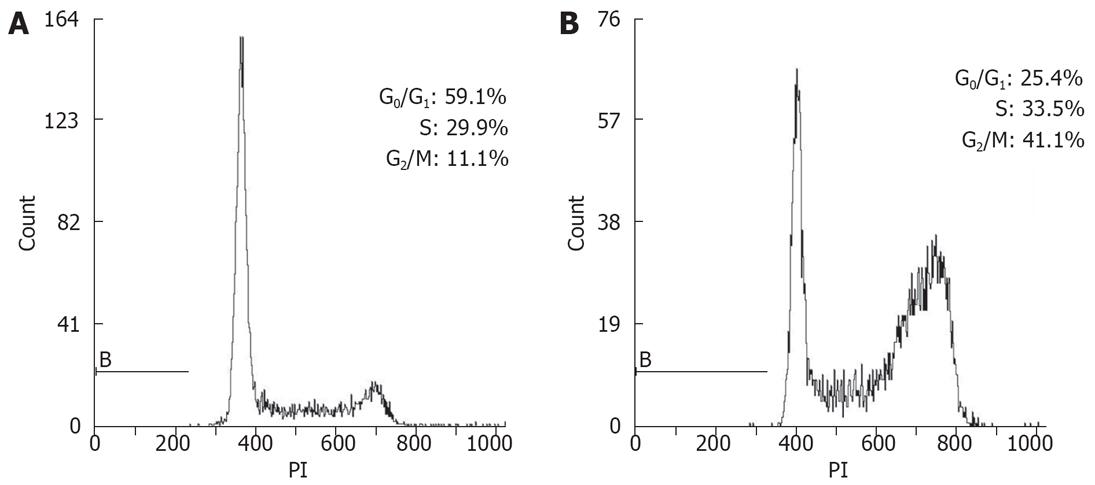
Figure 3 Lithium inhibited the proli-feration of Eca-109 cells by an arrest in G2/M phase.
Eca-109 cells were treated with lithium (20 mmol/L) for 0 h (A) and 24 h (B). Eca-109 cells were harvested and cell cycle profiles were obtained by staining with propidium iodide (PI).
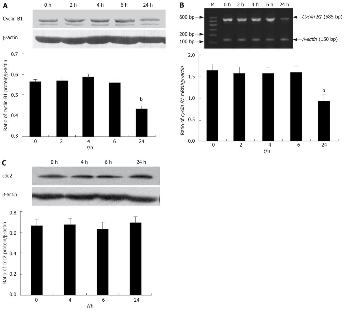
Figure 4 Effects of lithium on cell cycle regulatory molecules.
A: Western blotting analysis for cyclin B1 protein levels after treatment with 20 mmol/L lithium; B: RT-PCR analysis for quantification of the relative mRNA abundance of cyclin B1 after treatment with 20 mmol/L lithium; C: Western blotting analysis for cdc2 protein levels after treatment with 20 mmol/L lithium. bP < 0.01 vs the control group.
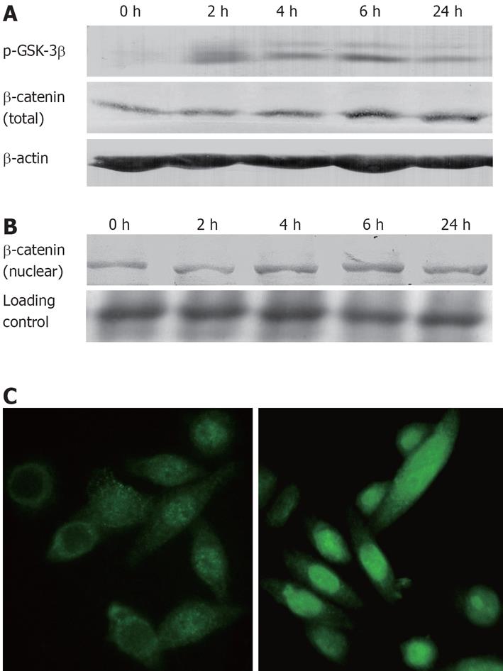
Figure 5 Lithium induced β-catenin stabilization via inhibition of GSK-3βin vitro.
A: Eca-109 cells were incubated with 20 mmol/L lithium for 0-24 h. Expressions of p-GSK-3β and total β-catenin protein were analyzed by Western blotting. The untreated Eca-109 cells (0 h) served as negative control; B: Western blotting analysis for β-catenin protein level of nuclear lysates; C: Immunofluorescence demonstrated a predominant cytoplasm localization of β-catenin in the untreated cells (left) whereas after incubation with lithium (20 mmol/L) for 6 h, β-catenin was predominantly located in the nucleus of Eca-109 cells (right).
Figure 6 Lithium induced cyclin D1 stabilization via inhibition of GSK-3βin vitro.
Eca-109 cells were incubated with 20 mmol/L lithium for 0-24 h. Expression of cyclin D1 was analyzed by Western blotting. The untreated Eca-109 cells (0 h) served as negative control.
- Citation: Wang JS, Wang CL, Wen JF, Wang YJ, Hu YB, Ren HZ. Lithium inhibits proliferation of human esophageal cancer cell line Eca-109 by inducing a G2/M cell cycle arrest. World J Gastroenterol 2008; 14(25): 3982-3989
- URL: https://www.wjgnet.com/1007-9327/full/v14/i25/3982.htm
- DOI: https://dx.doi.org/10.3748/wjg.14.3982


