Copyright
©2008 The WJG Press and Baishideng.
World J Gastroenterol. Jun 28, 2008; 14(24): 3866-3871
Published online Jun 28, 2008. doi: 10.3748/wjg.14.3866
Published online Jun 28, 2008. doi: 10.3748/wjg.14.3866
Figure 1 Different immunohistochemical (IHC) staining patterns for β-catenin in colorectal carcinomas.
A: In normal colonic epithelium, β-catenin is predominantly expressed in the cell membrane; B: A medium-powered view of a colonic adenocarcinoma showing membranous and cytoplasmic expression of β-catenin; C: This case shows intense nuclear expression of β-catenin.
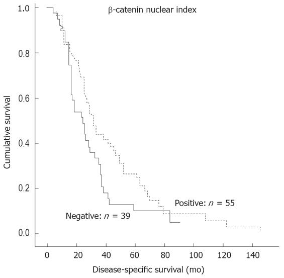
Figure 2 Disease-specific survival predicted by the nuclear index (NI) of colorectal tumors.
The stippled line: nuclear expression positive. The continuous line: nuclear expression negative. The Kaplan-Meier, log rank test; P = 0.046. One patient died of another cause and was excluded from analyses.
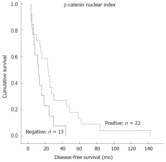
Figure 3 Disease-free survival predicted by the nuclear index (NI) of colorectal tumors.
The stippled line: nuclear expression positive. The continuous line: nuclear expression negative. The Kaplan-Meier, log rank test; P = 0.041.
- Citation: Elzagheid A, Buhmeida A, Korkeila E, Collan Y, Syrjänen K, Pyrhönen S. Nuclear β-catenin expression as a prognostic factor in advanced colorectal carcinoma. World J Gastroenterol 2008; 14(24): 3866-3871
- URL: https://www.wjgnet.com/1007-9327/full/v14/i24/3866.htm
- DOI: https://dx.doi.org/10.3748/wjg.14.3866


