Copyright
©2008 The WJG Press and Baishideng.
World J Gastroenterol. Jun 14, 2008; 14(22): 3591-3593
Published online Jun 14, 2008. doi: 10.3748/wjg.14.3591
Published online Jun 14, 2008. doi: 10.3748/wjg.14.3591
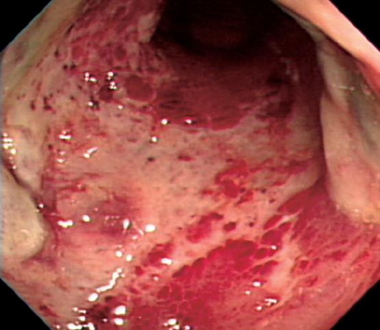
Figure 1 Sigmoidoscopy shows the severe erythematous friable mucosa and diffuse ulceration with dirty exudates.
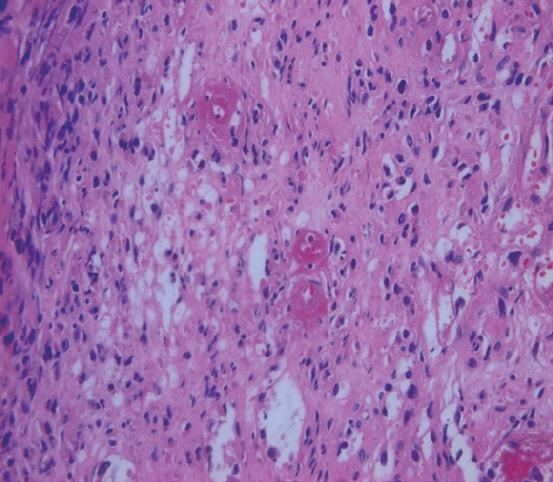
Figure 2 Microscopy of colon biopsies shows the thickening of small vessel wall and lymphocyte infiltration around vessels (HE, × 400).
- Citation: Lee JR, Paik CN, Kim JD, Chung WC, Lee KM, Yang JM. Ischemic colitis associated with intestinal vasculitis: Histological proof in systemic lupus erythematosus. World J Gastroenterol 2008; 14(22): 3591-3593
- URL: https://www.wjgnet.com/1007-9327/full/v14/i22/3591.htm
- DOI: https://dx.doi.org/10.3748/wjg.14.3591


