Copyright
©2008 The WJG Press and Baishideng.
World J Gastroenterol. Jun 14, 2008; 14(22): 3579-3582
Published online Jun 14, 2008. doi: 10.3748/wjg.14.3579
Published online Jun 14, 2008. doi: 10.3748/wjg.14.3579
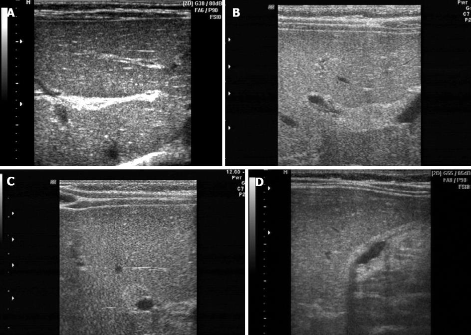
Figure 1 Ultrasonography.
A: Fibrous plaque of inversed triangular shape at the confluent site of right and left hepatic ducts; B: Longitudinal section of streak-shaped fibrous plaque at the confluent site of right and left hepatic ducts; C: Cross section of streak-shaped fibrous plaque at hepatic portal vein (arrow indicates fibrous plaque surrounding the right anterior area of the right branch of portal vein); D: Flat and small gallbladder with low tension. The thickness of capsule wall was uneven.
- Citation: Li SX, Zhang Y, Sun M, Shi B, Xu ZY, Huang Y, Mao ZQ. Ultrasonic diagnosis of biliary atresia: A retrospective analysis of 20 patients. World J Gastroenterol 2008; 14(22): 3579-3582
- URL: https://www.wjgnet.com/1007-9327/full/v14/i22/3579.htm
- DOI: https://dx.doi.org/10.3748/wjg.14.3579


