Copyright
©2008 The WJG Press and Baishideng.
World J Gastroenterol. Jun 14, 2008; 14(22): 3511-3517
Published online Jun 14, 2008. doi: 10.3748/wjg.14.3511
Published online Jun 14, 2008. doi: 10.3748/wjg.14.3511
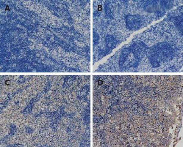
Figure 1 Tissue microarrays of mesenteric lymph nodes were prepared and stained for mmunohistochemistry (× 200).
(A) NF-κB expression in the treated group (6 h); (B) Bax protein expression in the sham-operated group (3 h); (C) Bax protein expression in the treated group (12 h); (D) Bcl-2 protein expression in the treated group (3 h).
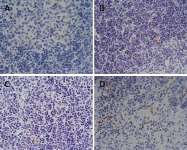
Figure 2 TUNEL staining, showing the pathological changes in the mesenteric lymph nodes (× 400).
(A) In the sham-operated group (6 h), there were no apoptotic cells; (B) In the model group (12 h), several apoptotic cells appeared; (C) treated group (6 h); (D) apoptotic cells were increased in the treated group (12 h).
- Citation: Zhang XP, Xu HM, Jiang YY, Yu S, Cai Y, Lu B, Xie Q, Ju TF. Influence of dexamethasone on mesenteric lymph node of rats with severe acute pancreatitis. World J Gastroenterol 2008; 14(22): 3511-3517
- URL: https://www.wjgnet.com/1007-9327/full/v14/i22/3511.htm
- DOI: https://dx.doi.org/10.3748/wjg.14.3511


