Copyright
©2008 The WJG Press and Baishideng.
World J Gastroenterol. May 28, 2008; 14(20): 3273-3275
Published online May 28, 2008. doi: 10.3748/wjg.14.3273
Published online May 28, 2008. doi: 10.3748/wjg.14.3273
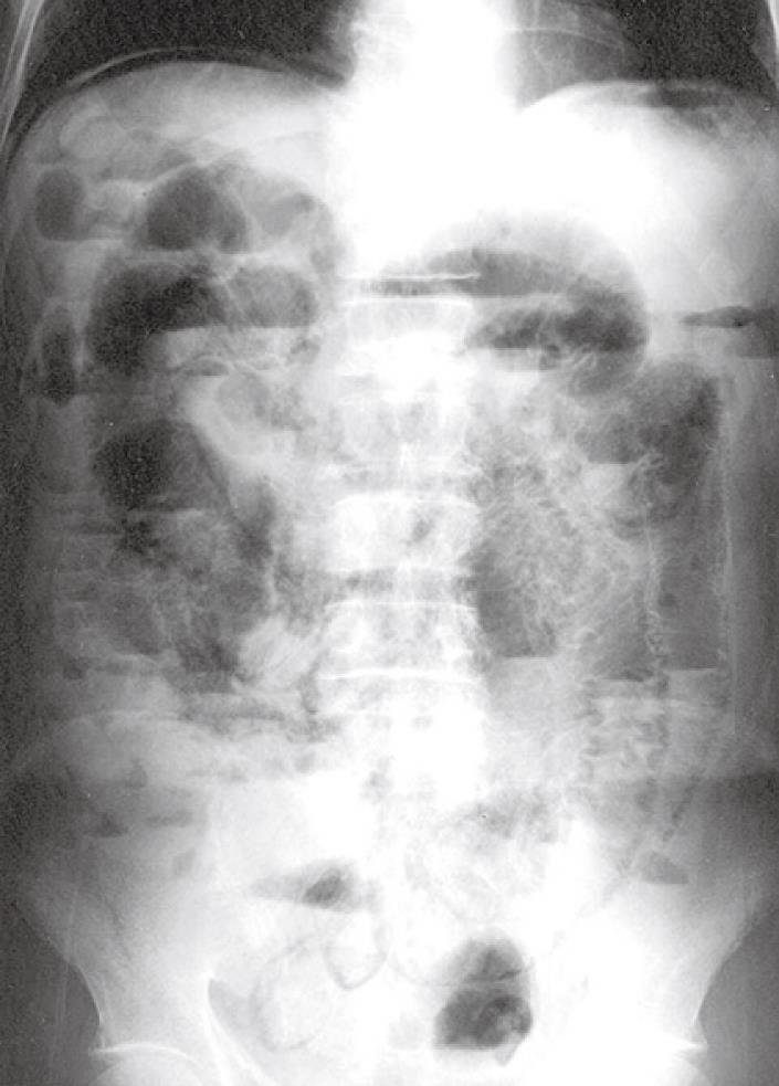
Figure 1 Abdominal radiogram showing intraluminal gases in the entire small intestine and free air under the diaphragm.
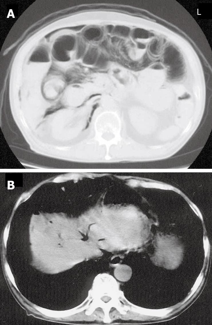
Figure 2 Abdominal CT scan showing excessive intraluminal gases in the entire small intestine and free air in the retroperitoneal space (A), and free air in the falciform ligament (B).
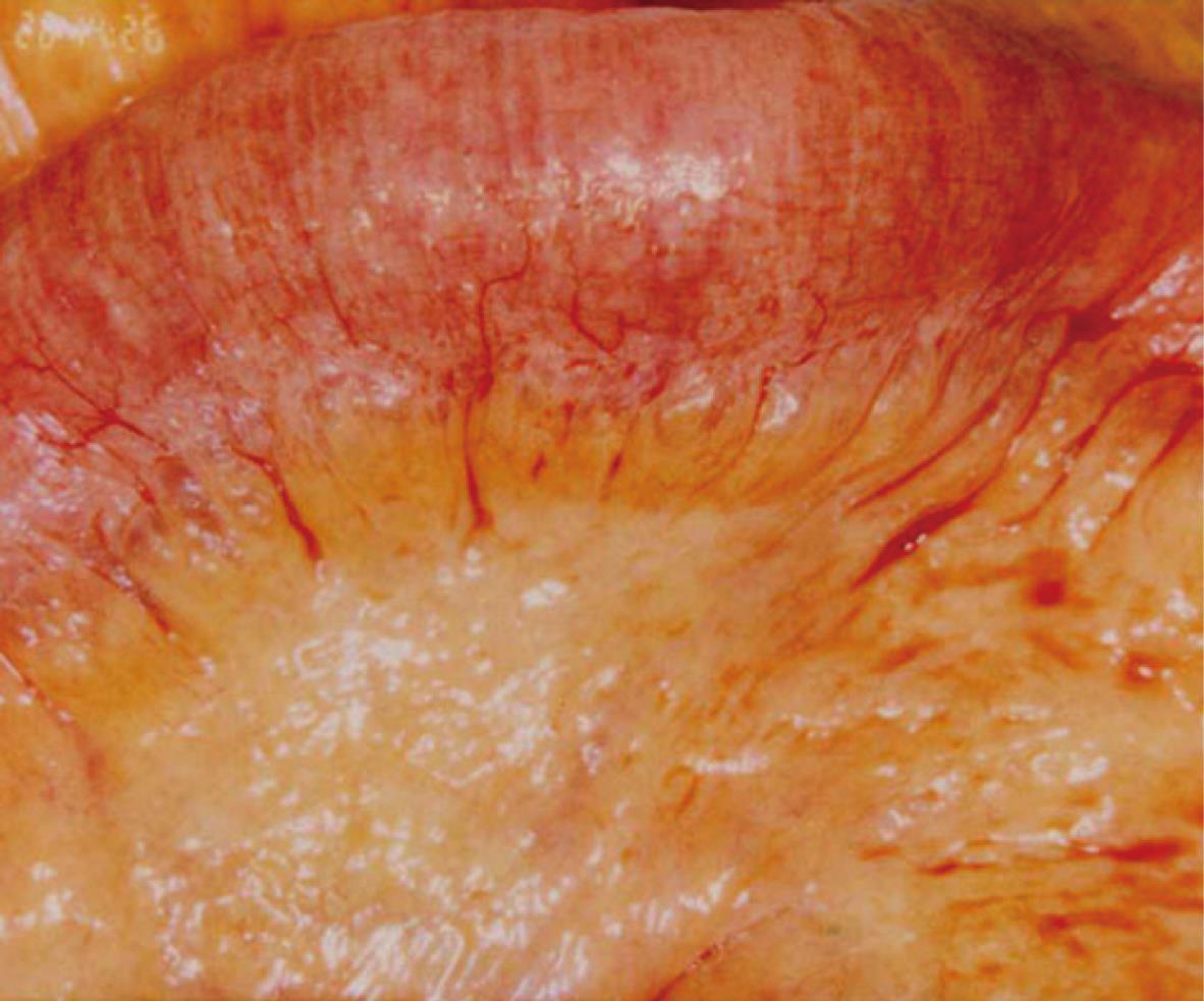
Figure 3 Expanded intraluminal air spaces in the small intestine and mesenterium during intra-operation.
- Citation: Mimatsu K, Oida T, Kawasaki A, Kano H, Kuboi Y, Aramaki O, Amano S. Pneumatosis cystoides intestinalis after fluorouracil chemotherapy for rectal cancer. World J Gastroenterol 2008; 14(20): 3273-3275
- URL: https://www.wjgnet.com/1007-9327/full/v14/i20/3273.htm
- DOI: https://dx.doi.org/10.3748/wjg.14.3273


