Copyright
©2008 The WJG Press and Baishideng.
World J Gastroenterol. May 28, 2008; 14(20): 3262-3265
Published online May 28, 2008. doi: 10.3748/wjg.14.3262
Published online May 28, 2008. doi: 10.3748/wjg.14.3262
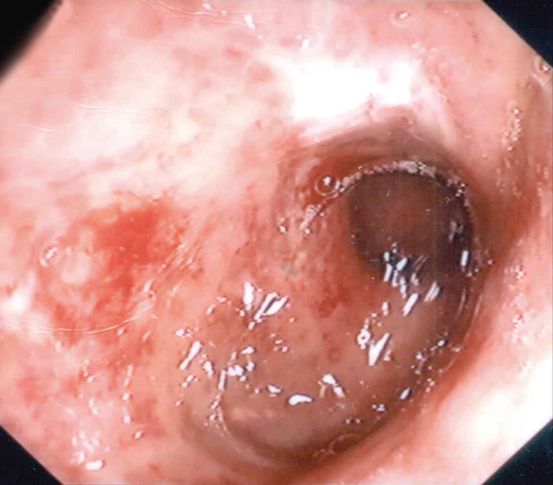
Figure 1 Endoscopic image of patient’s ischemic colitis.
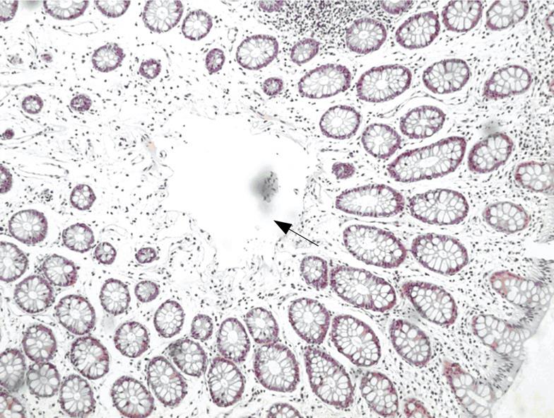
Figure 2 ΗΕ staining of colonic mucosa with an air bubble in the lamina propria (arrow).
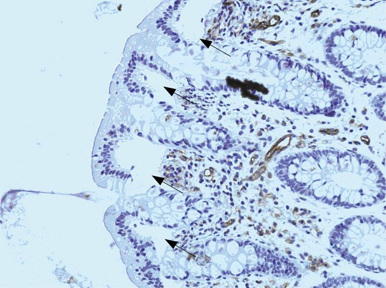
Figure 3 Immunohistochemistry for CD31 revealing lack of staining in the areas of air bubbles in the lamina propria (arrows).
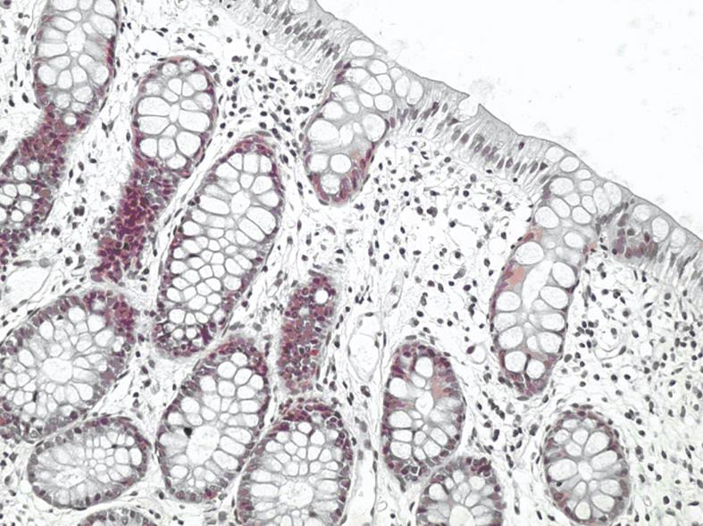
Figure 4 HE staining of normal histologic appearance of our patient’s colonic mucosa, 2 mo after the ischemic colitis episode.
- Citation: Goumas K, Poulou A, Tyrmpas I, Dandakis D, Bartzokis S, Tsamouri M, Barbati K, Soutos D. Acute ischemic colitis during scuba diving: Report of a unique case. World J Gastroenterol 2008; 14(20): 3262-3265
- URL: https://www.wjgnet.com/1007-9327/full/v14/i20/3262.htm
- DOI: https://dx.doi.org/10.3748/wjg.14.3262


