Copyright
©2008 The WJG Press and Baishideng.
World J Gastroenterol. May 14, 2008; 14(18): 2939-2941
Published online May 14, 2008. doi: 10.3748/wjg.14.2939
Published online May 14, 2008. doi: 10.3748/wjg.14.2939
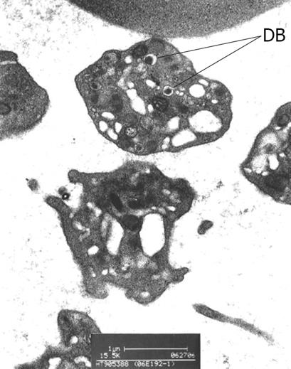
Figure 1 Electron photomicrograph of the patient’s plaletets, showing a diminished number (normal, 4-8) of dense bodies (DB) in one, and absence of dense bodies in the adjacent platelets.
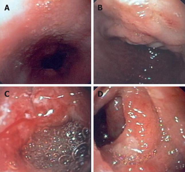
Figure 2 Pictures obtained at esophagogastroduodenoscopy.
A: Esophagus appeared normal except for mild nodularis near gastroesophageal junction; B: Stomach was covered with a coffee-ground substance and a patch of mucosal irregularity at proximal lesser curve was seen; C: A solitary ulcer of 2 cm in diameter with a rolled margin was found at anterior wall of the first part of duodenum; D: Multiple small, flat and oval aphthoid ulcers were found in the second part of duodenum.
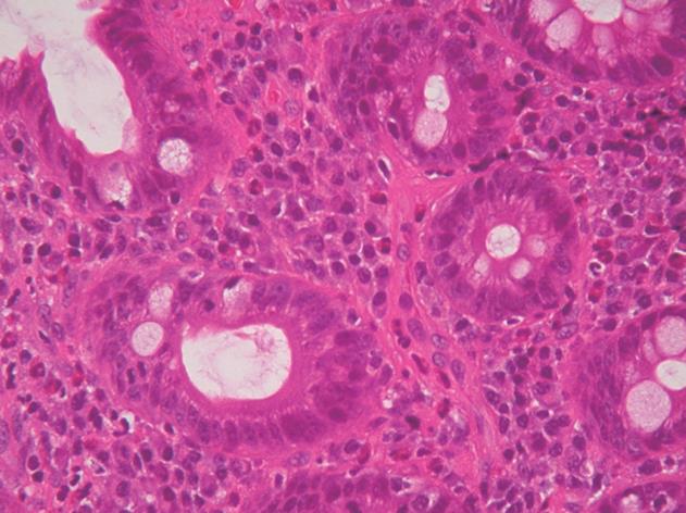
Figure 3 Histologic section of duodenal mucosa showing intact villous architecture with a moderate inflammatory infiltrate of lymphocytes, plasma cells and eosinophils in lamina propria.
- Citation: Lee ACW, Poon KH, Lo WH, Wong LG. Chronic ulcerative gastroduodenitis as a first gastrointestinal manifestation of Hermansky-Pudlak syndrome in a 10-year-old child. World J Gastroenterol 2008; 14(18): 2939-2941
- URL: https://www.wjgnet.com/1007-9327/full/v14/i18/2939.htm
- DOI: https://dx.doi.org/10.3748/wjg.14.2939


