Copyright
©2008 The WJG Press and Baishideng.
World J Gastroenterol. May 14, 2008; 14(18): 2932-2934
Published online May 14, 2008. doi: 10.3748/wjg.14.2932
Published online May 14, 2008. doi: 10.3748/wjg.14.2932
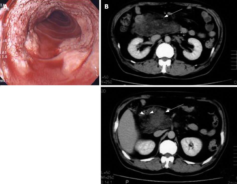
Figure 1 A: Gastrointestinal endoscopy revealed bleeding at the duodenum; B: Computed tomography demonstrating a heterogenous mass (arrow) at the pancreatic head and suspected invasion to the duodenal wall (arrowhead).
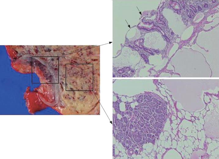
Figure 2 Histology consists of a benign soft tissue mass with lymphatic and blood vessels.
Arrows show invasion to the duodenal wall.
- Citation: Toyoki Y, Hakamada K, Narumi S, Nara M, Kudoh D, Ishido K, Sasaki M. A case of invasive hemolymphangioma of the pancreas. World J Gastroenterol 2008; 14(18): 2932-2934
- URL: https://www.wjgnet.com/1007-9327/full/v14/i18/2932.htm
- DOI: https://dx.doi.org/10.3748/wjg.14.2932


