Copyright
©2008 The WJG Press and Baishideng.
World J Gastroenterol. May 14, 2008; 14(18): 2928-2931
Published online May 14, 2008. doi: 10.3748/wjg.14.2928
Published online May 14, 2008. doi: 10.3748/wjg.14.2928
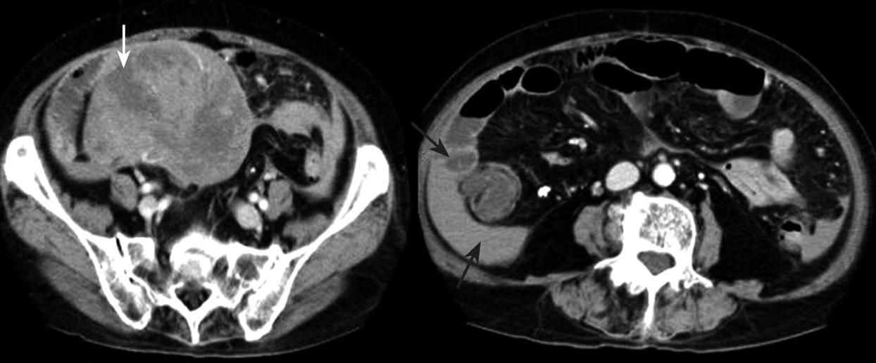
Figure 1 CT scan demonstrating a large heterogeneous mass with a non-uniform enhancement pattern (white arrow) in the pelvis and hemoperitoneum (black arrows).
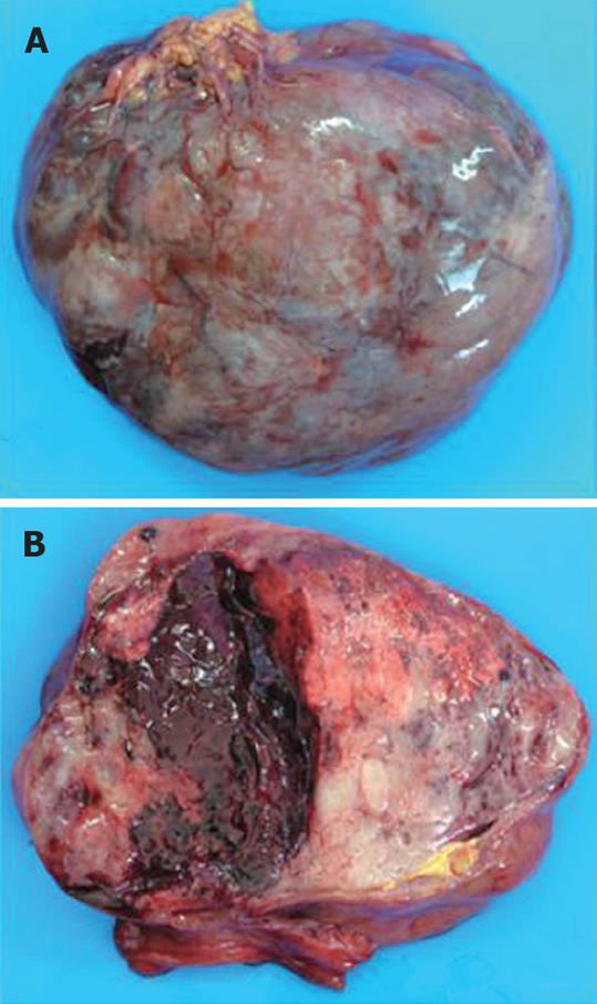
Figure 2 Macroscopic finding of the tumor.
A: A large tumor (measuring 13 cm × 11 cm) arising from the ileum with extraluminal growth; B: The cut surface showing bleeding blood clots in the tumor.
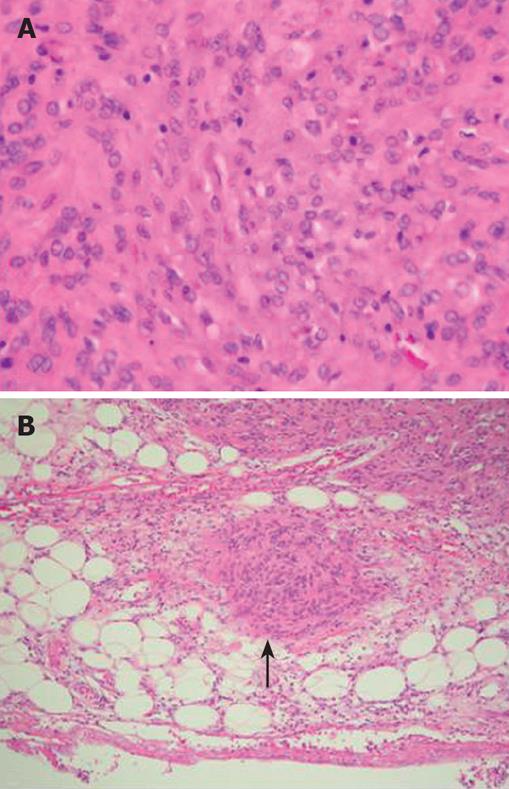
Figure 3 Microscopic findings of the tumor.
A: Histological examination demonstrating interlaced bundles of large Bizarre spindle cells without mitotic figures (HE, × 100); B: Tumor cells present in the subserosa (black arrow) (HE, × 20).
- Citation: Hirasaki S, Fujita K, Matsubara M, Kanzaki H, Yamane H, Okuda M, Suzuki S, Shirakawa A, Saeki H. A ruptured large extraluminal ileal gastrointestinal stromal tumor causing hemoperitoneum. World J Gastroenterol 2008; 14(18): 2928-2931
- URL: https://www.wjgnet.com/1007-9327/full/v14/i18/2928.htm
- DOI: https://dx.doi.org/10.3748/wjg.14.2928


