Copyright
©2008 The WJG Press and Baishideng.
World J Gastroenterol. May 14, 2008; 14(18): 2917-2919
Published online May 14, 2008. doi: 10.3748/wjg.14.2917
Published online May 14, 2008. doi: 10.3748/wjg.14.2917
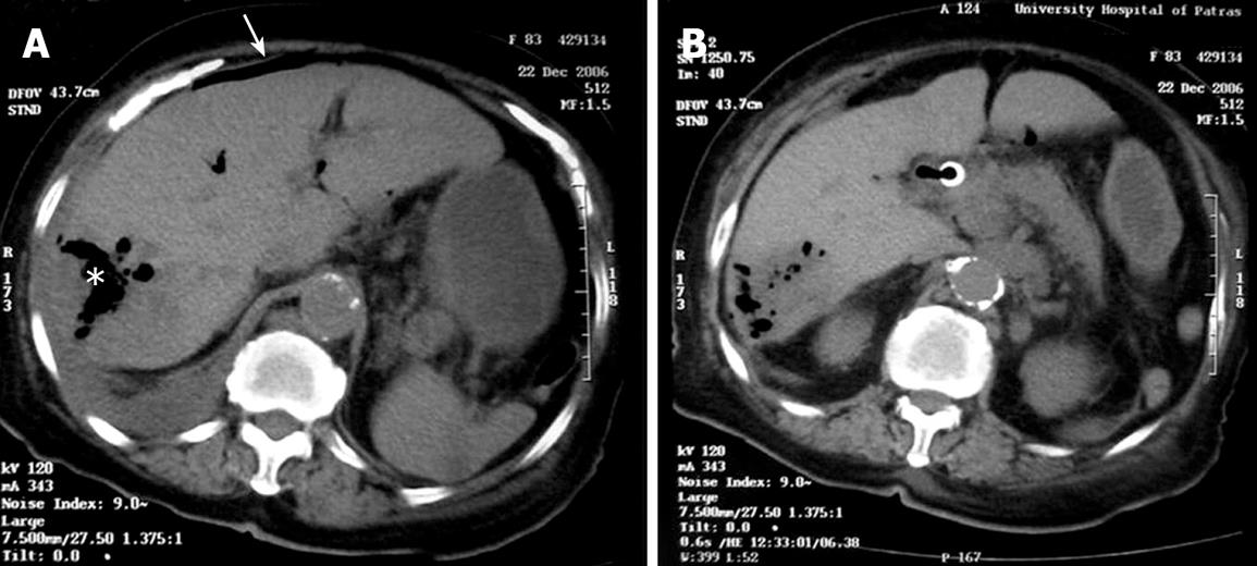
Figure 1 Contrast-enhanced abdominal CT showing an extensive necrotic air-containing lesion touching Glisson’s capsule in the posterior right hepatic lobe within a metastatic hepatic mass (asterisk) and free air (arrow) (A).
The metallic stent in the common bile duct is at the proper position and there is pneumobilia of the common bile duct and intrahepatic bile ducts of the metastatic mass in the posterior right hepatic lobe (B).
- Citation: Assimakopoulos SF, Thomopoulos KC, Giali S, Triantos C, Siagris D, Gogos C. A rare etiology of post-endoscopic retrograde cholangiopancreatography pneumoperitoneum. World J Gastroenterol 2008; 14(18): 2917-2919
- URL: https://www.wjgnet.com/1007-9327/full/v14/i18/2917.htm
- DOI: https://dx.doi.org/10.3748/wjg.14.2917


