Copyright
©2008 The WJG Press and Baishideng.
World J Gastroenterol. Apr 28, 2008; 14(16): 2586-2589
Published online Apr 28, 2008. doi: 10.3748/wjg.14.2586
Published online Apr 28, 2008. doi: 10.3748/wjg.14.2586
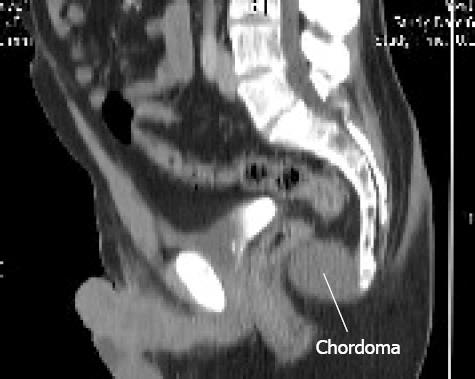
Figure 1 Post processing sagittal reconstruction of non-contrast pelvic CT demonstrating a hypo attenuating spherical lesion anterior to the coccyx.
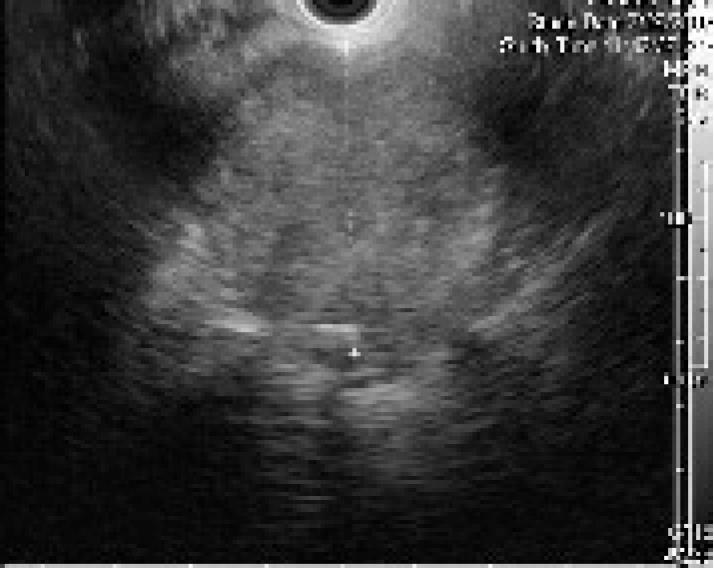
Figure 2 Endoscopic ultrasound with the Olympus GF-UM160 radial echoendoscope demonstrates a well circumscribed hyperechoic lesion, with some focal areas of heterogeneous echotexture, corresponding to the lesion described in Figure 1.
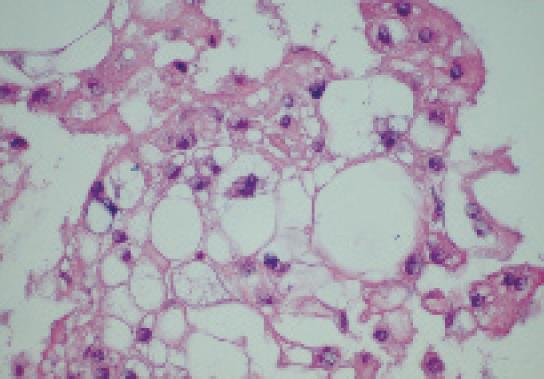
Figure 3 Tissue section of the aspirate sample shows sheets of vacuolated "physaliphorous" cells, classic of chordoma.
These cells demonstrate immunorea-ctivity with pan-cytokeratin and epithelial membrane antigen stains.
- Citation: Gottlieb K, Lin PH, Liu DM, Anders K. Transrectal EUS-guided FNA biopsy of a presacral chordoma-report of a case and review of the literature. World J Gastroenterol 2008; 14(16): 2586-2589
- URL: https://www.wjgnet.com/1007-9327/full/v14/i16/2586.htm
- DOI: https://dx.doi.org/10.3748/wjg.14.2586


