Copyright
©2008 The WJG Press and Baishideng.
World J Gastroenterol. Apr 28, 2008; 14(16): 2566-2571
Published online Apr 28, 2008. doi: 10.3748/wjg.14.2566
Published online Apr 28, 2008. doi: 10.3748/wjg.14.2566
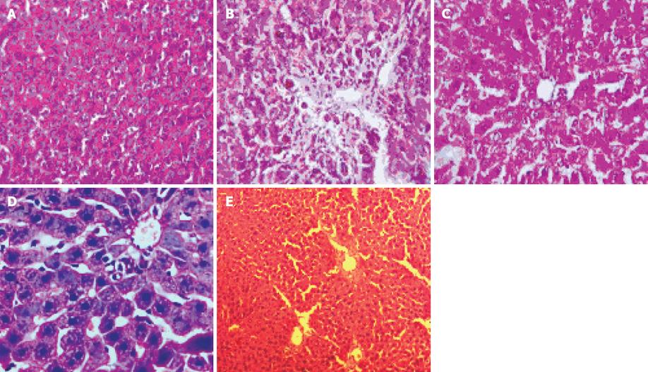
Figure 1 Photograph of rat liver shows (HE, × 100).
A: Liver of a control rat showing normal hepatocytes and normal architecture; B: Liver section from a CCl4 treated rat demonstrating the destruction of architectural pattern, nodule formation in the lobular zone, inflamed periportal zone, moderate inflammation of portal area; C: Liver section from a Silymarin treated rat showing regeneration of normal hepatocytes; D: Liver section from a R. emodi treated rat showing normal lobular architecture; E: Liver section from a S. mukorossi treated rat showing normal lobular architecture no necrosis or fatty changes or any inflammatory reaction can be seen.
-
Citation: Ibrahim M, Khaja MN, Aara A, Khan AA, Habeeb MA, Devi YP, Narasu ML, Habibullah CM. Hepatoprotective activity of
Sapindus mukorossi andRheum emodi extracts:In vitro andin vivo studies. World J Gastroenterol 2008; 14(16): 2566-2571 - URL: https://www.wjgnet.com/1007-9327/full/v14/i16/2566.htm
- DOI: https://dx.doi.org/10.3748/wjg.14.2566


