Copyright
©2008 The WJG Press and Baishideng.
World J Gastroenterol. Apr 14, 2008; 14(14): 2277-2279
Published online Apr 14, 2008. doi: 10.3748/wjg.14.2277
Published online Apr 14, 2008. doi: 10.3748/wjg.14.2277
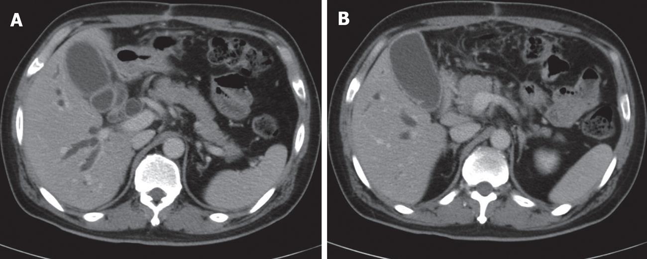
Figure 1 Abdominal CT scan findings.
A: Dilated intra- and extrahepatic bile ducts; B: Abrupt cut-off of the distal common bile duct.
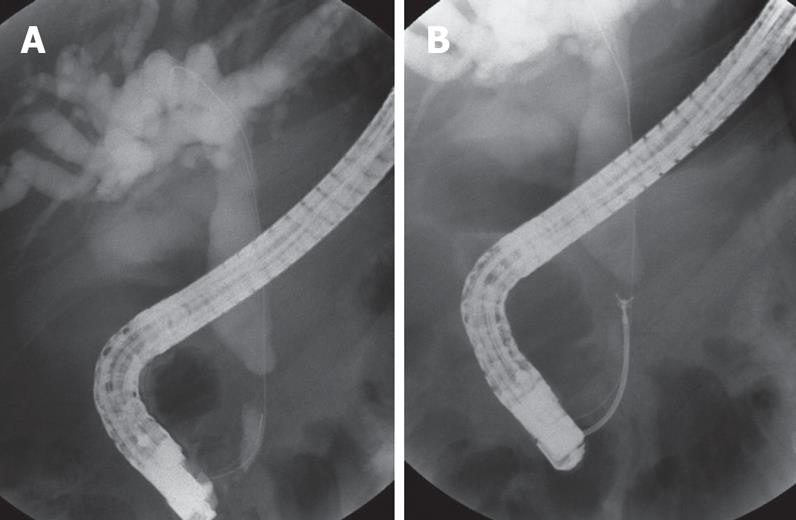
Figure 2 ERCP findings.
A: Stenosis of the distal CBD, approximately 1 cm in length, located just above the pancreas, with ductal dilatation above the narrowed portion; B: Biopsy of the stricture was obtained.
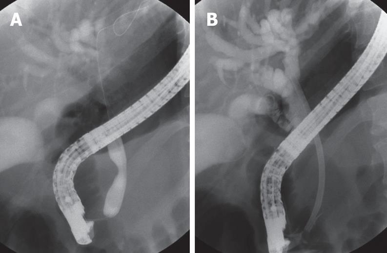
Figure 3 ERCP findings.
A: The CBD was dilated with TTS balloons, but the dilatation was not effective; B: An Amsterdam-type plastic stent (12Fr, 7 cm) was placed across the stricture.
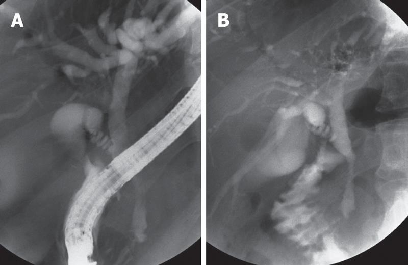
Figure 4 Follow-up ERCP findings.
A: Improvement of the CBD stricture; B: There is normal passage of radiocontrast material into the bile duct through the papilla.
- Citation: Kang DO, Kim TH, You SS, Min HJ, Kim HJ, Jung WT, Lee OJ. Successful endoscopic treatment of biliary stricture following mesenteric tear caused by blunt abdominal trauma. World J Gastroenterol 2008; 14(14): 2277-2279
- URL: https://www.wjgnet.com/1007-9327/full/v14/i14/2277.htm
- DOI: https://dx.doi.org/10.3748/wjg.14.2277


