Copyright
©2008 The WJG Press and Baishideng.
World J Gastroenterol. Apr 7, 2008; 14(13): 2121-2123
Published online Apr 7, 2008. doi: 10.3748/wjg.14.2121
Published online Apr 7, 2008. doi: 10.3748/wjg.14.2121
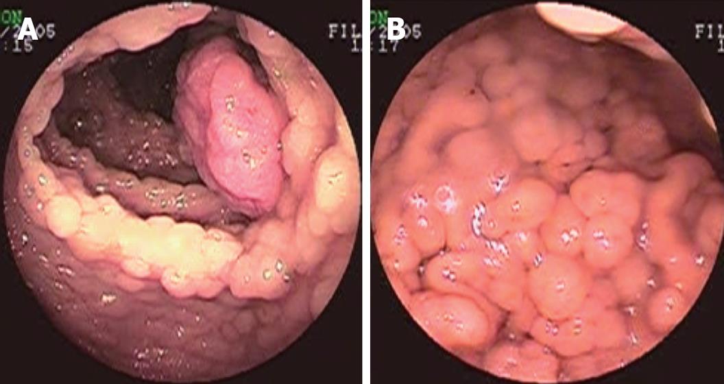
Figure 1 Preoperative endoscopy examination.
A: Colonoscopy showing multiple polyps covering the entire colon and rectum; B: Gastroscopy showing multiple polyps covering the fundus and corpus ventriculi.
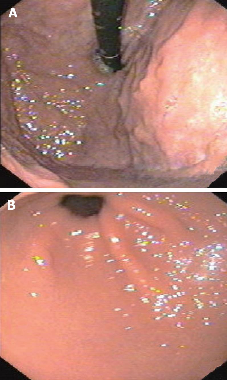
Figure 2 Postoperative gastroscopy examination shows disappearance of polyps of the fundus and corpus ventriculi (A) and smooth mucous membrane of the corpus ventriculi and pars pylorica (B).
- Citation: Gu GL, Wang SL, Wei XM, Bai L. Diagnosis and treatment of Gardner syndrome with gastric polyposis: A case report and review of the literature. World J Gastroenterol 2008; 14(13): 2121-2123
- URL: https://www.wjgnet.com/1007-9327/full/v14/i13/2121.htm
- DOI: https://dx.doi.org/10.3748/wjg.14.2121


