Copyright
©2008 The WJG Press and Baishideng.
World J Gastroenterol. Apr 7, 2008; 14(13): 2118-2120
Published online Apr 7, 2008. doi: 10.3748/wjg.14.2118
Published online Apr 7, 2008. doi: 10.3748/wjg.14.2118
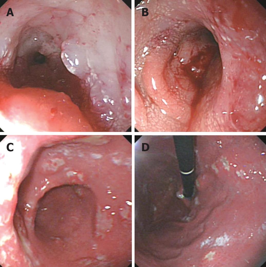
Figure 1 Colonoscopic pictures (A) and (B) show stenosing primary rectal tumour with multiple ulcerations; Gastroscopy (C) and (D) showed multiple ulcers in the stomach.
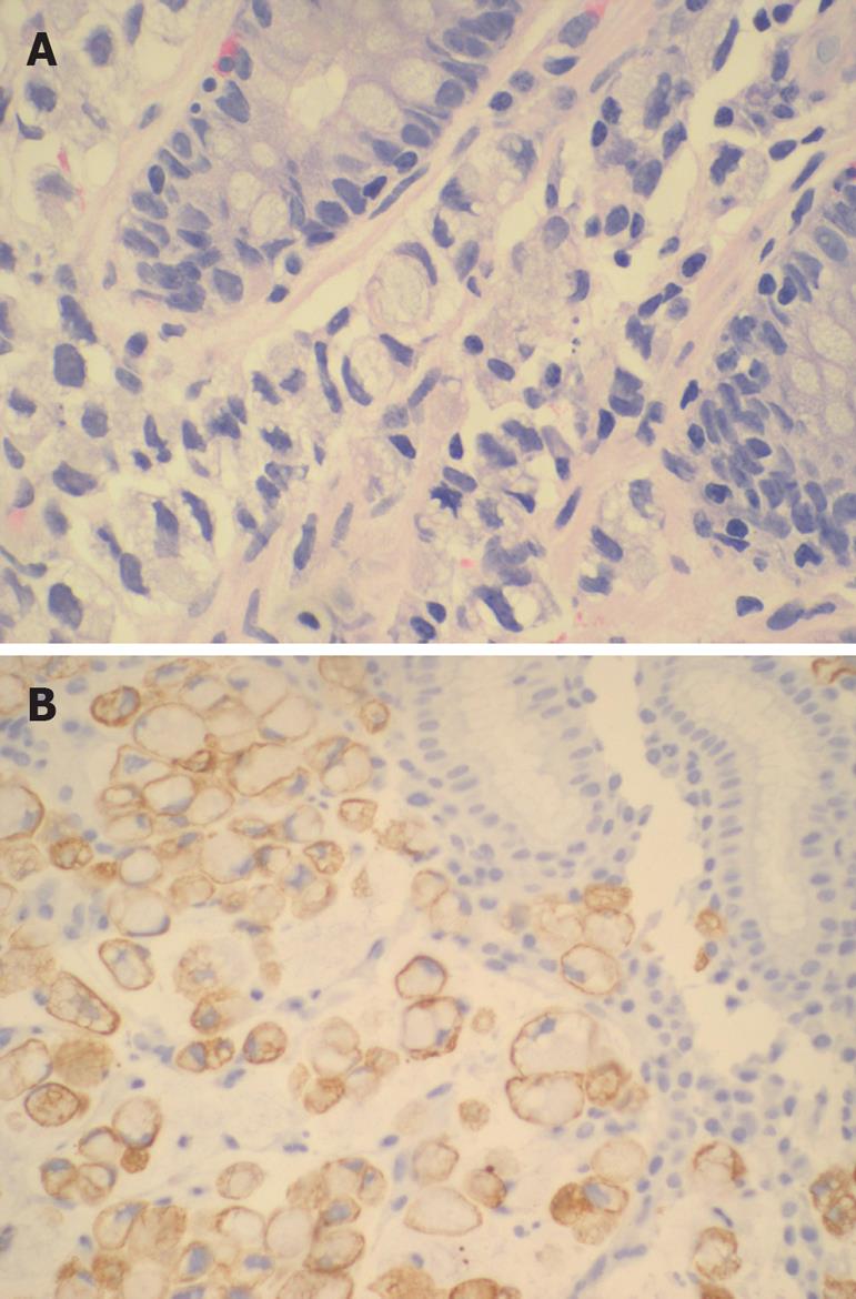
Figure 2 Photomicrograph A: normal colonic glands with signet ring cells within the lamina propria (HE); B: gastric mucosa with similar signet ring cells which were positive for CK 7.
- Citation: Sim HL, Tan KY, Poon PL, Cheng A. Primary rectal signet ring cell carcinoma with peritoneal dissemination and gastric secondaries. World J Gastroenterol 2008; 14(13): 2118-2120
- URL: https://www.wjgnet.com/1007-9327/full/v14/i13/2118.htm
- DOI: https://dx.doi.org/10.3748/wjg.14.2118


