Copyright
©2008 The WJG Press and Baishideng.
World J Gastroenterol. Mar 21, 2008; 14(11): 1797-1799
Published online Mar 21, 2008. doi: 10.3748/wjg.14.1797
Published online Mar 21, 2008. doi: 10.3748/wjg.14.1797
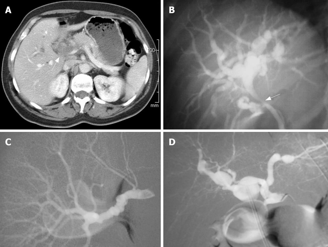
Figure 1 Clinical examination results.
A: Abdominal CT scan showing a soft tissue mass encapsulating the portal vein and hepatic artery (arrow); B: ERCP showing a common hepatic duct stricture (arrow) with proximal biliary tree dilatation; C: Selective angiography of the renal artery demonstrating the classic appearance of “string of beads”; D: Selective angiography of the hepatic artery revealing multiple aneurysms.
- Citation: Shussman N, Edden Y, Mintz Y, Verstandig A, Rivkind AI. Hemobilia due to hepatic artery aneurysm as the presenting sign of fibro-muscular dysplasia. World J Gastroenterol 2008; 14(11): 1797-1799
- URL: https://www.wjgnet.com/1007-9327/full/v14/i11/1797.htm
- DOI: https://dx.doi.org/10.3748/wjg.14.1797


