Copyright
©2008 The WJG Press and Baishideng.
World J Gastroenterol. Mar 14, 2008; 14(10): 1603-1611
Published online Mar 14, 2008. doi: 10.3748/wjg.14.1603
Published online Mar 14, 2008. doi: 10.3748/wjg.14.1603
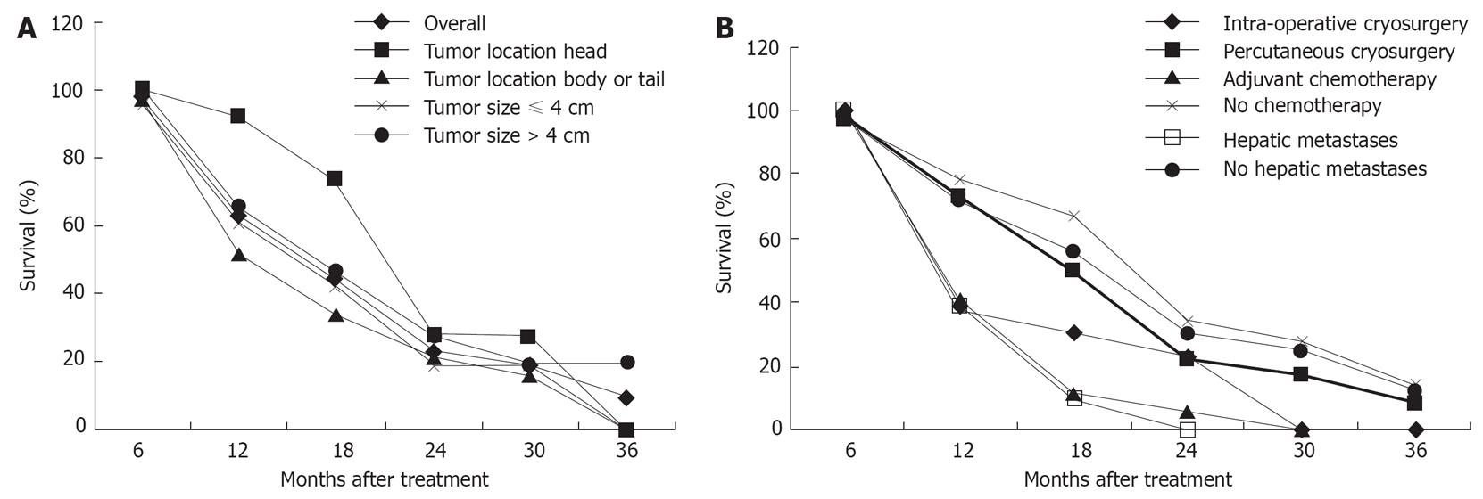
Figure 1 Survival rates of patients with pancreatic cancer.
A: Overall results and those in patients with different location and size of the tumor; B: Pancreatic cancer with different modes of therapies, and with or without hepatic metastases.
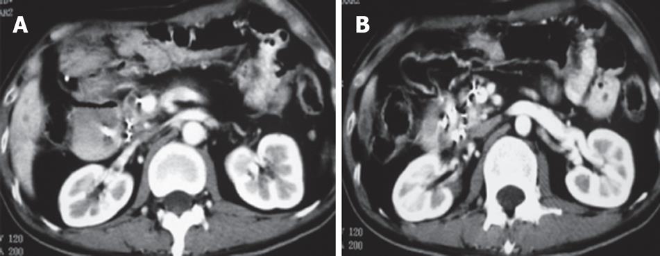
Figure 2 Pancreatic CT scan in case 1.
A: Before treatment; B: Three months after treatment; C: Twelve months after treatment.
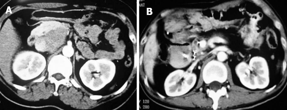
Figure 3 CT scan of case 2.
A: Before treatment, a mass was seen in pancreatic body and trail; B: 0ne month after treatment; C: Six months after treatment.
Figure 4 CT scan in case 3.
A: One months after treatment; B: Three months after treatment.
Figure 5 CT scan in case 4.
A: Before treatment; B: Eight months after treatment.
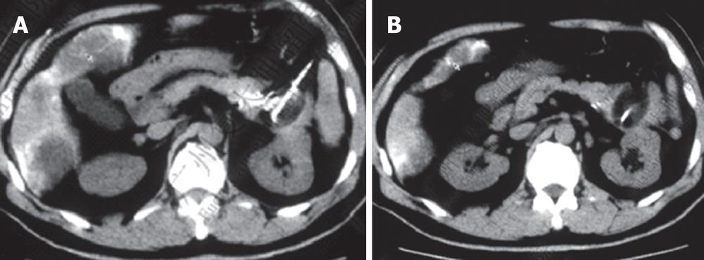
Figure 6 CT scan in case 5.
A and B showing cryoprobe and 125iodine seeds in different layers during treatment.
Figure 7 CT and PET-CT in case 6.
A: CT at 10 mo after the first treatment, showing 125iodine seeds; B: PET-CT at 12 mo after the first treatment showing new lesion in pancreatic body; C: PET-CT at 3 mo after second treatment.
- Citation: Xu KC, Niu LZ, Hu YZ, He WB, He YS, Li YF, Zuo JS. A pilot study on combination of cryosurgery and 125iodine seed implantation for treatment of locally advanced pancreatic cancer. World J Gastroenterol 2008; 14(10): 1603-1611
- URL: https://www.wjgnet.com/1007-9327/full/v14/i10/1603.htm
- DOI: https://dx.doi.org/10.3748/wjg.14.1603


