Copyright
©2008 The WJG Press and Baishideng.
World J Gastroenterol. Mar 14, 2008; 14(10): 1528-1533
Published online Mar 14, 2008. doi: 10.3748/wjg.14.1528
Published online Mar 14, 2008. doi: 10.3748/wjg.14.1528
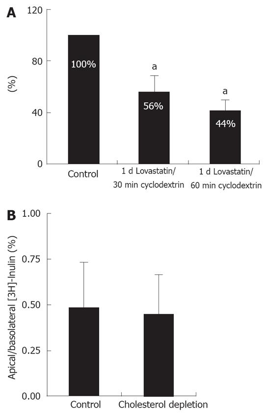
Figure 1 Cholesterol can be depleted from Caco-2 cells by a combination of lovastatin and MβCD.
Cells were grown for 1d in the presence of lovastatin/ mevalonate and then treated for 30-60 min with 10 mmol/L MβCD. A: Depending on the time of MβCD extraction, the total cellular cholesterol levels could be reduced by about 60%. Exposure for 30min with 10 mmol/L MβCD (2nd bar) was used for the following experiments. The cholesterol level was arbitrarily set to 100% in control cells. An asterisk indicates significant differences (56.3% ± 17.1% vs 100% and 46.3% ± 17.1% vs 100%, both aP < 0.05); B: [3H]-inulin permeability did not change after cholesterol depletion (P > 0.05).
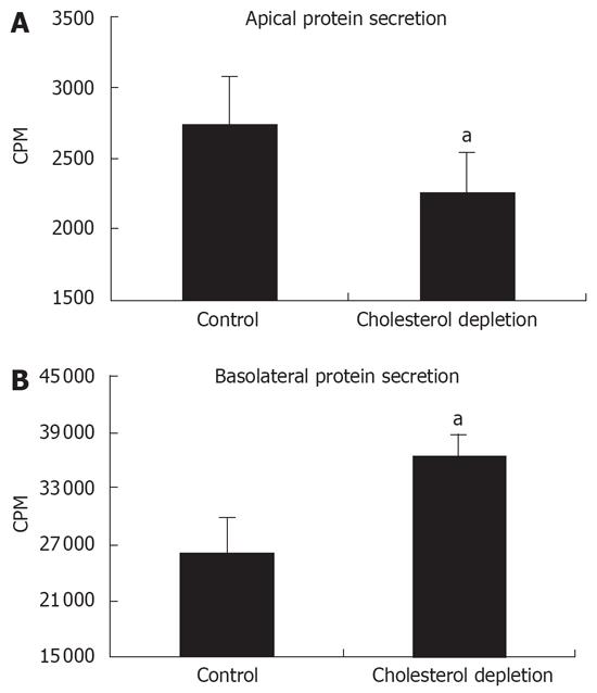
Figure 2 Effect of cholesterol depletion on polarized protein secretion.
Filter-grown Caco-2 cells were pulsed for 1 h with [35S]-methionine and the secretion of labeled proteins within 2 h was quantified using a scintillation counter. The total radioactivity of protein precipitates was (A) reduced in the apical medium of cholesterol depleted cells and (B) increased in the basolateral medium. An asterisk indicates significant differences (2736.6 ± 352.7 vs 2257.6 ± 282.4 and 28 893.2 ± 3523.1 vs 38 256.0 ± 2555.3, aP < 0.05).
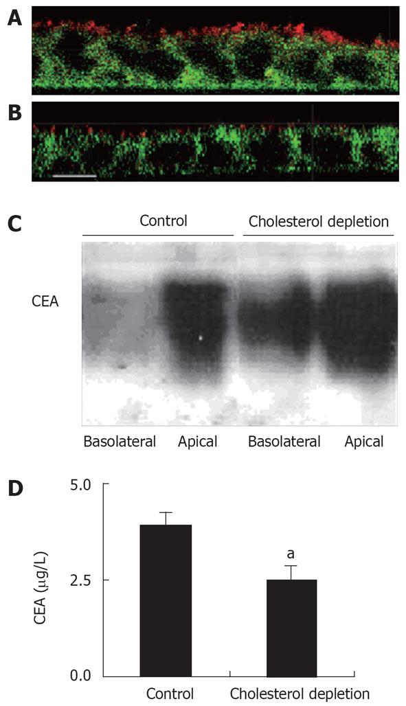
Figure 3 The effect of cholesterol depletion on apical and basolateral secretion of CEA.
A, B: X-Z confocal views of cells labeled with antibodies directed against CEA (red) and Na+/K+-ATPase (green). In non-depleted cells under steady state conditions, CEA family proteins are expressed at the apical surface (A). Caco-2 cells were grown to confluency on polycarbonate filters for 14 d and cholesterol-depleted with a combination of lovastatin/MβCD. After cholesterol depletion, CEA was still apical but with a lower expression level (B) but no basolateral staining of CEA was detected (Bar 10 &mgr;m). C: Media from apical and basolateral chamber were collected for 2 h and Western blots performed. In non-depleted cells, most of the CEA is secreted into the apical medium. In cholesterol depleted cells, a significant amount was also found in the basolateral medium. D: Quantification of apical CEA secretion using an ECLISA from Roche. Six similar experiments were performed. An asterisk indicates significant differences (3.12 ± 0.62 vs 1.91 ± 0.81, aP < 0.05).
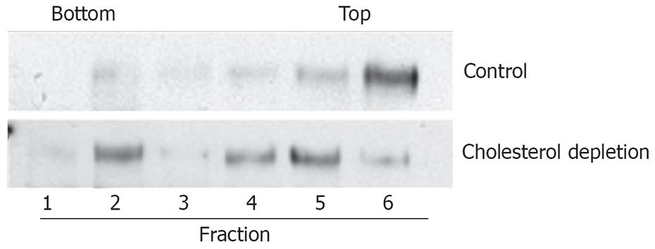
Figure 4 Cholesterol depletion reduces association of CEA with DRMs.
Filter grown Caco-2 cells with or without treatment with a combination of lovastatin/MβCD were extracted on ice with 2% Triton X-100. After flotation in an OptiPrep step-gradient, fractions were collected and Western blots performed. Cholesterol depletion shifted CEA from the top (raft fractions) to the bottom fractions (non raft fractions).
- Citation: Ehehalt R, Krautter M, Zorn M, Sparla R, Füllekrug J, Kulaksiz H, Stremmel W. Increased basolateral sorting of carcinoembryonic antigen in a polarized colon carcinoma cell line after cholesterol depletion-Implications for treatment of inflammatory bowel disease. World J Gastroenterol 2008; 14(10): 1528-1533
- URL: https://www.wjgnet.com/1007-9327/full/v14/i10/1528.htm
- DOI: https://dx.doi.org/10.3748/wjg.14.1528


