Copyright
©2008 The WJG Press and Baishideng.
World J Gastroenterol. Jan 7, 2008; 14(1): 152-154
Published online Jan 7, 2008. doi: 10.3748/wjg.14.152
Published online Jan 7, 2008. doi: 10.3748/wjg.14.152
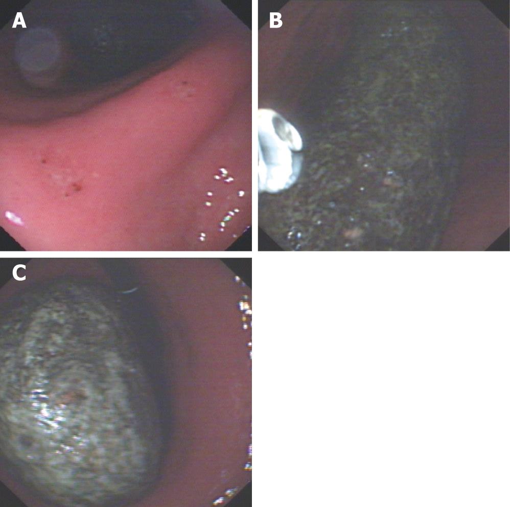
Figure 1 A: A huge disopyrobezoar within the stomach shown by gastroscopy, with two erosions at the corner of stomach; B and C: A huge ellipse disopyrobezoar shown gastroscopically within the stomach.
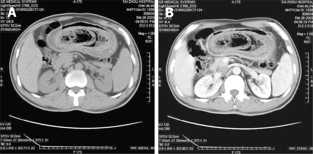
Figure 2 A: Unenhanced CT scan showing a huge gastric mass with air bubbles and mottled appearance; B: Enhanced CT scan showing the same image as demonstrated on the unenhanced CT scan.
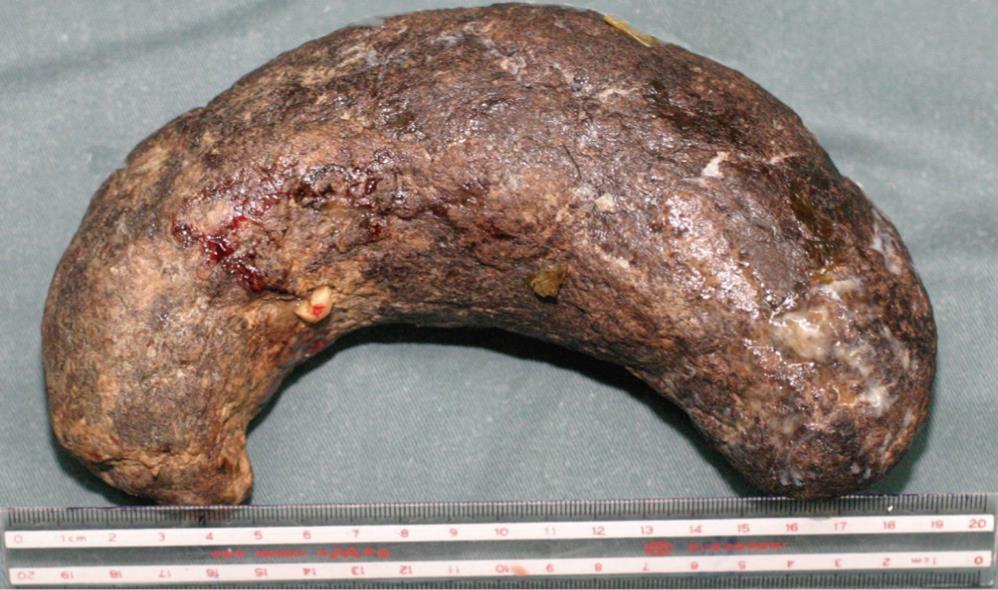
Figure 3 Surgical specimen of gastric disopyrobezoar,18 cm long, 7.
5 cm in diameter, with visible brownish persimmon remnants on its surface.
- Citation: Zhang RL, Yang ZL, Fan BG. Huge gastric disopyrobezoar: A case report and review of literatures. World J Gastroenterol 2008; 14(1): 152-154
- URL: https://www.wjgnet.com/1007-9327/full/v14/i1/152.htm
- DOI: https://dx.doi.org/10.3748/wjg.14.152


