Copyright
©2008 The WJG Press and Baishideng.
World J Gastroenterol. Jan 7, 2008; 14(1): 146-151
Published online Jan 7, 2008. doi: 10.3748/wjg.14.146
Published online Jan 7, 2008. doi: 10.3748/wjg.14.146
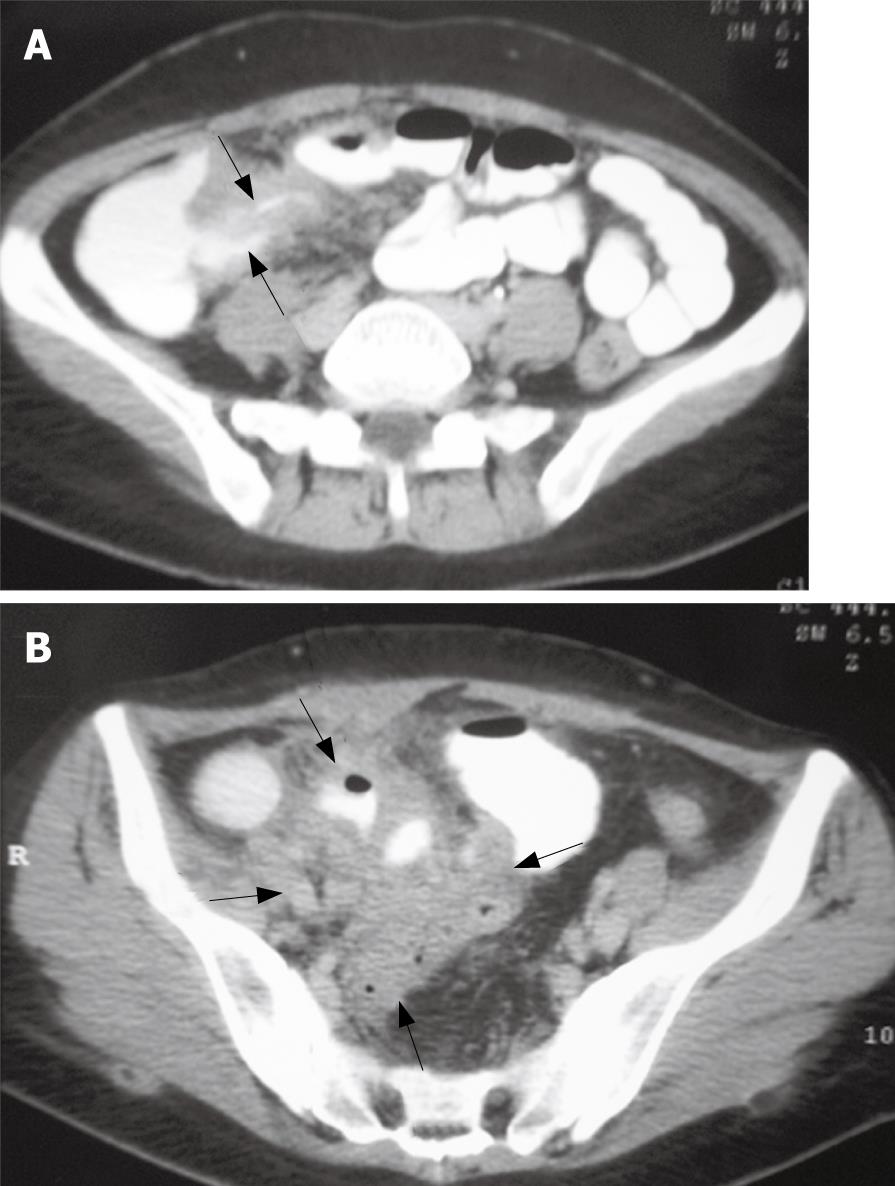
Figure 1 Contrast-enhanced scan showing the mural thickening of terminal ileum (arrows) (A), and a complex, predominantly inflammatory mass of large size (6.
4 cm × 6.1 cm) (arrows) (B).
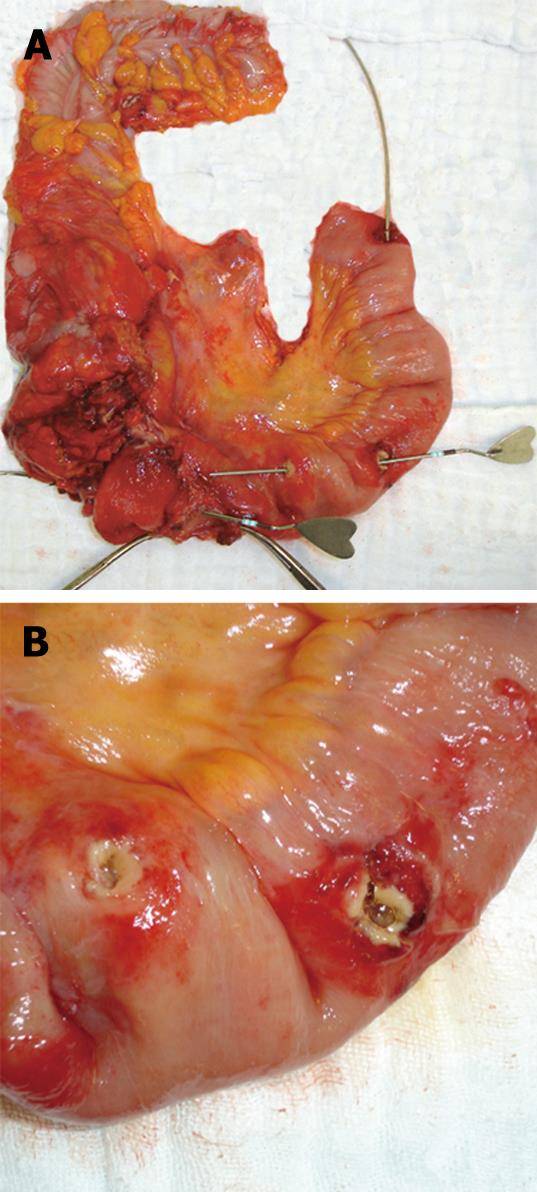
Figure 2 Gross appearance of the resected specimen showing Crohn’s ileitis with multiple fistulas probed with instruments (A), and ileal segment with two adjacent openings of an internal enteric fistula after separation of adhesions (B).
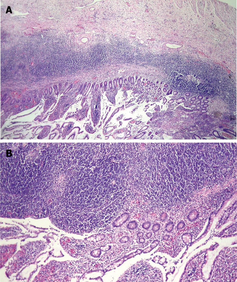
Figure 3 Microscopic examination of the resected specimen revealed transmural inflammatory cell infiltration with crypt distortion (A), and transmural lymphoid aggregates (B) (HE, A × 10, B × 20).
- Citation: Teke Z, Aytekin FO, Atalay AO, Demirkan NC. Crohn’s disease complicated by multiple stenoses and internal fistulas clinically mimicking small bowel endometriosis. World J Gastroenterol 2008; 14(1): 146-151
- URL: https://www.wjgnet.com/1007-9327/full/v14/i1/146.htm
- DOI: https://dx.doi.org/10.3748/wjg.14.146


