Copyright
©2007 Baishideng Publishing Group Co.
World J Gastroenterol. Mar 7, 2007; 13(9): 1347-1351
Published online Mar 7, 2007. doi: 10.3748/wjg.v13.i9.1347
Published online Mar 7, 2007. doi: 10.3748/wjg.v13.i9.1347
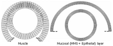
Figure 1 The configurations of the separated muscle (left) and the mucosal (right) layer at the zero-stress state (open sectors) and the no-load state (closed circular rings).
The no-load state was obtained by forcing the opening sector to be closed using a pure bending deformation. The pure bending loads are indicted by arrows. The numerical model was conducted by using the ABAQUS software.
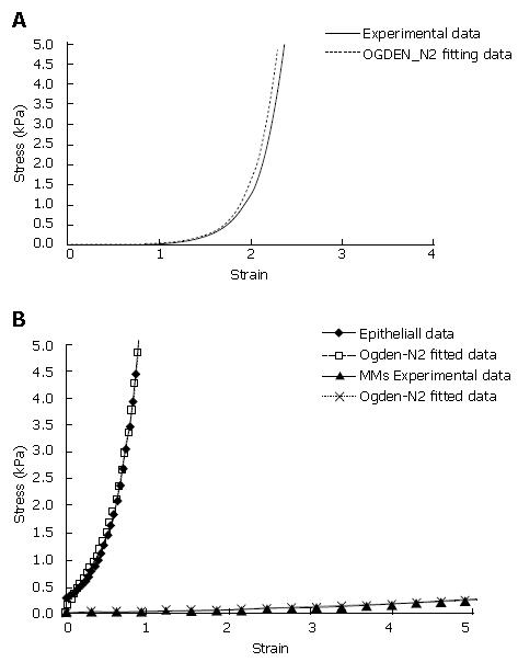
Figure 2 Circumferential experimental and curvefitted stress-strain curves for the muscle (A) and mucosal (B) layers.
The curves were fitted by using the Ogden 2nd order strain energy function. The stress-strain curve of the epithelial layer was tested from the mucosal-submucosal layer while the stress strain curve of the MMS layer was assumed to have a similar pattern as the epithelial layer with the magnitude about one order softer than the epithelial layer. For the circumferential uni-axial test and the circumferential planar test the separated muscle and mucosal-submucosal rings were fixed in two L shape rods, one end of the L shape rod was connected to a cannula. The cannulas were connected to rods that could be moved at controlled velocities by a motor. One of the rods was attached to a force transducer. The distance between the rods was adjusted manually to the in vitro length of the strips (reference length). The force (F) was recorded online by a Labview program.
Math 3 Math(A3).
Math 4 Math(A4).
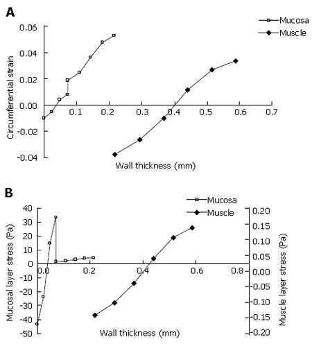
Figure 3 The circumferential strain (A) and stress (B) distribution throughout the muscle and mucosal (MMS + epithelial) layers after the separated muscle and mucosal layer sectors were closed.
The strain and stress jump occurred at the interface between the epithelial and MMS layer.
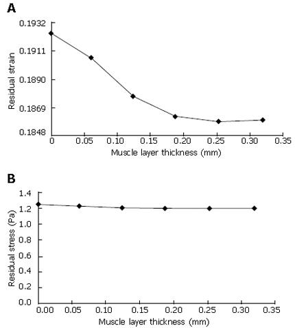
Figure 4 Circumferential residual strain (A) and residual Mises stress (B) distribution in the muscle layer at the no-load state.
Strain was positive throughout the muscle wall.
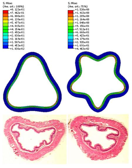
Figure 5 Residual stress of the mucosal layer in a three folds model (Top, left) and five folds model (Top, right).
The morphological oesophageal images indicated that the folds numbers are between three (bottom, left) and five (bottom, right) at the no-load state.
- Citation: Liao D, Zhao J, Yang J, Gregersen H. The oesophageal zero-stress state and mucosal folding from a GIOME perspective. World J Gastroenterol 2007; 13(9): 1347-1351
- URL: https://www.wjgnet.com/1007-9327/full/v13/i9/1347.htm
- DOI: https://dx.doi.org/10.3748/wjg.v13.i9.1347


