Copyright
©2007 Baishideng Publishing Group Co.
World J Gastroenterol. Feb 28, 2007; 13(8): 1295-1298
Published online Feb 28, 2007. doi: 10.3748/wjg.v13.i8.1295
Published online Feb 28, 2007. doi: 10.3748/wjg.v13.i8.1295
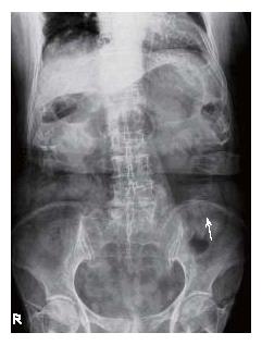
Figure 1 Plain abdominal film showing a vague stone not identified in the left iliac fossa (arrow).
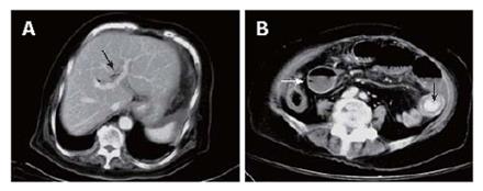
Figure 2 Abdominal computed tomography scan showing air in the biliary tree (arrow) (A) and an ectopic stone (arrow) impacting the jejunal lumen accompanying dilatation of proximal small bowel loops (white arrow) (B).
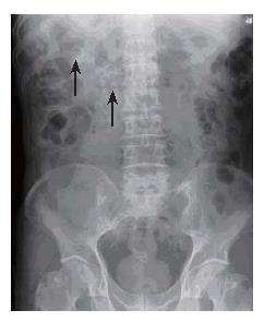
Figure 3 Plain abdominal film showing two round calcified stones in the right upper quadrant of abdomen (arrow).
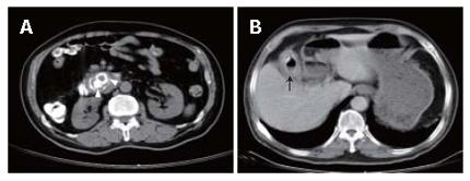
Figure 4 Abdominal computed tomography scan showing two round calcified stones impacting the proximal third portion of the duodenum (arrowhead) (A) and a thickened wall gallbladder with internal air and small calcified content (arrow) (B).
- Citation: Chou JW, Hsu CH, Liao KF, Lai HC, Cheng KS, Peng CY, Yang MD, Chen YF. Gallstone ileus: Report of two cases and review of the literature. World J Gastroenterol 2007; 13(8): 1295-1298
- URL: https://www.wjgnet.com/1007-9327/full/v13/i8/1295.htm
- DOI: https://dx.doi.org/10.3748/wjg.v13.i8.1295


