Copyright
©2007 Baishideng Publishing Group Co.
World J Gastroenterol. Feb 21, 2007; 13(7): 1135-1137
Published online Feb 21, 2007. doi: 10.3748/wjg.v13.i7.1135
Published online Feb 21, 2007. doi: 10.3748/wjg.v13.i7.1135
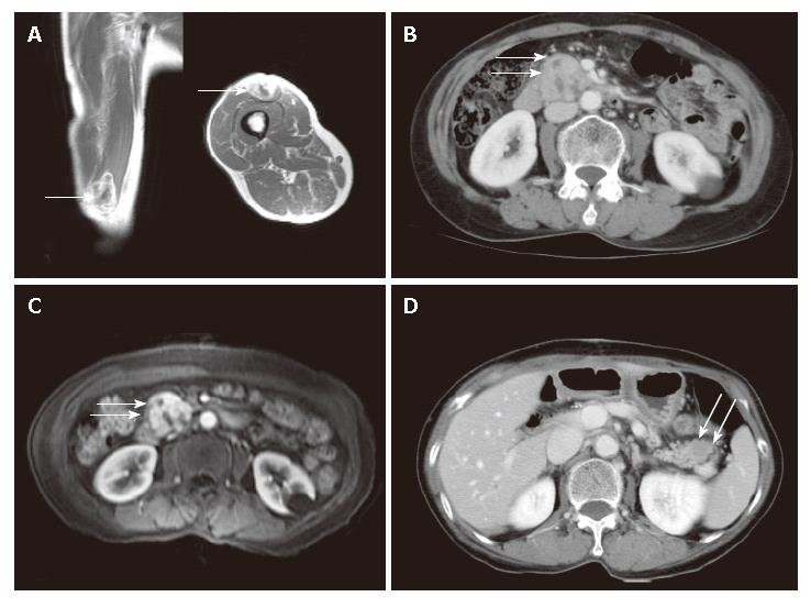
Figure 1 Radiological findings.
A: Gadolinium-enhanced T1 weighted image showing a soft tissue tumor in the muscle of the anterior thigh; B: Contrast-enhanced abdominal CT scan showing a heterogeneously enhanced mass at the head of the pancreas; C: Gadolinium-enhanced T1 weighted image showing a well-enhanced mass with necrotic foci at the head of the pancreas; D: Follow-up CT obtained after 10 mo showing a low attenuated mass in the tail of the pancreas.
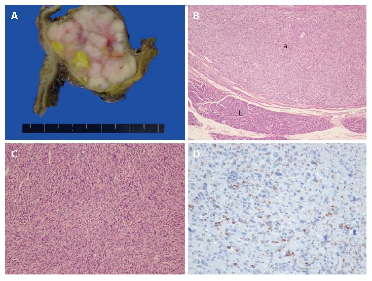
Figure 2 Gross and microscopic findings for metastatic leiomyo-sarcoma.
A: The section reveals a whitish solid tumor with a lobulated pattern and a yellowish necrotic portion, abutting duodenal wall on the left side, and compressed pancreatic head tissue on the right side; B: Low-power view displaying eosinophilic intersecting fascicles circumscribed by a fibrous capsule (a) and adjacent normal pancreatic tissue (b) (hematoxylin and eosin stain ×40); C: Cellular proliferation of atypical spindle tumor cells is accompanied by nuclear atypia and hyperchromasia (hematoxylin and eosin stain × 200); D: Tumor cells show immunoreactivity for desmin.
- Citation: Koh YS, Chul J, Cho CK, Kim HJ. Pancreatic metastasis of leiomyosarcoma in the right thigh: A case report. World J Gastroenterol 2007; 13(7): 1135-1137
- URL: https://www.wjgnet.com/1007-9327/full/v13/i7/1135.htm
- DOI: https://dx.doi.org/10.3748/wjg.v13.i7.1135


