Copyright
©2007 Baishideng Publishing Group Co.
World J Gastroenterol. Feb 21, 2007; 13(7): 1085-1089
Published online Feb 21, 2007. doi: 10.3748/wjg.v13.i7.1085
Published online Feb 21, 2007. doi: 10.3748/wjg.v13.i7.1085
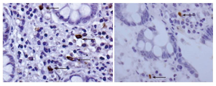
Figure 1 Immunohistochemical staining for HCMV of biopsy samples from ileoanal pouch.
Cells in the submucosa (arrows) show strong nuclear staining. Original magnification x 600.
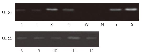
Figure 2 Agarose gel showing UL32 gene products (lanes 1-4, 5 and 6) and UL 55 gene products (lanes 8-12).
W: Water; N: HCMV-negative patient.
- Citation: Casadesus D, Tani T, Wakai T, Maruyama S, Iiai T, Okamoto H, Hatakeyama K. Possible role of human cytomegalovirus in pouchitis after proctocolectomy with ileal pouch-anal anastomosis in patients with ulcerative colitis. World J Gastroenterol 2007; 13(7): 1085-1089
- URL: https://www.wjgnet.com/1007-9327/full/v13/i7/1085.htm
- DOI: https://dx.doi.org/10.3748/wjg.v13.i7.1085


