Copyright
©2007 Baishideng Publishing Group Co.
World J Gastroenterol. Feb 7, 2007; 13(5): 804-805
Published online Feb 7, 2007. doi: 10.3748/wjg.v13.i5.804
Published online Feb 7, 2007. doi: 10.3748/wjg.v13.i5.804
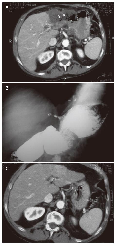
Figure 1 Enhanced CT abdominal scan performed after the percutaneous procedure showing air inclusions in the bile ducts within the left portion of the liver (A), radiograph of the first gastrointestinal tract showing a biliary gastric fistula starting from the lesser curvature of the stomach with concomitant spasm of the greater curvature (B), and enhanced CT scan of the abdomen performed before percutaneous thermal ablation of the HCC lesion in the III liver segment showing a left portion of the liver without aerobilia (C).
- Citation: Falco A, Orlando D, Sciarra R, Sergiacomo L. A case of biliary gastric fistula following percutaneous radiofrequency thermal ablation of hepatocellular carcinoma. World J Gastroenterol 2007; 13(5): 804-805
- URL: https://www.wjgnet.com/1007-9327/full/v13/i5/804.htm
- DOI: https://dx.doi.org/10.3748/wjg.v13.i5.804


