Copyright
©2007 Baishideng Publishing Group Co.
World J Gastroenterol. Feb 7, 2007; 13(5): 801-803
Published online Feb 7, 2007. doi: 10.3748/wjg.v13.i5.801
Published online Feb 7, 2007. doi: 10.3748/wjg.v13.i5.801
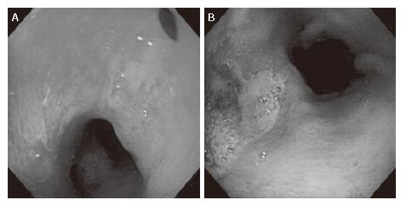
Figure 1 Upper gastrointestinal endoscopy showing a fistula in the esophagus, 20 mm proximal to the anastomosis (A), and a gastric ulcer just below the anastomosis (B).
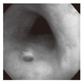
Figure 2 Bronchoscopic examination showing a fistula in the membranous portion of the trachea, 90 mm distal to the vocal cord.
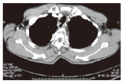
Figure 3 Computed tomography showing an esophagotracheal fistula located on a level with the upper wedge of sternum.
- Citation: Maruyama K, Motoyama S, Okuyama M, Sato Y, Hayashi K, Minamiya Y, Ogawa JI. Esophagotracheal fistula caused by gastroesophageal reflux 9 years after esophagectomy. World J Gastroenterol 2007; 13(5): 801-803
- URL: https://www.wjgnet.com/1007-9327/full/v13/i5/801.htm
- DOI: https://dx.doi.org/10.3748/wjg.v13.i5.801


