Copyright
©2007 Baishideng Publishing Group Inc.
World J Gastroenterol. Dec 21, 2007; 13(47): 6321-6326
Published online Dec 21, 2007. doi: 10.3748/wjg.v13.i47.6321
Published online Dec 21, 2007. doi: 10.3748/wjg.v13.i47.6321
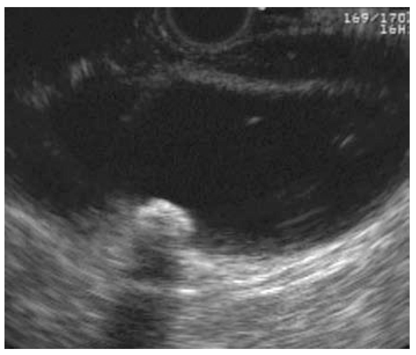
Figure 1 Linear EUS of gallbladder with shadowing stone.
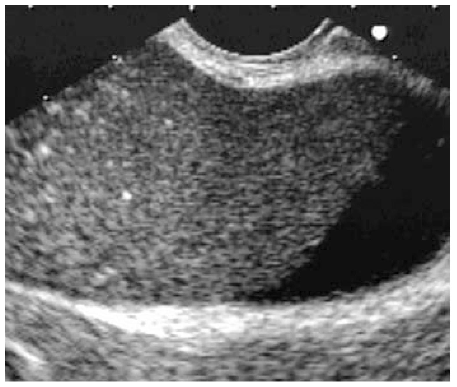
Figure 2 Linear EUS of gallbladder with sludge, and bright shadowing foci representing small stones.
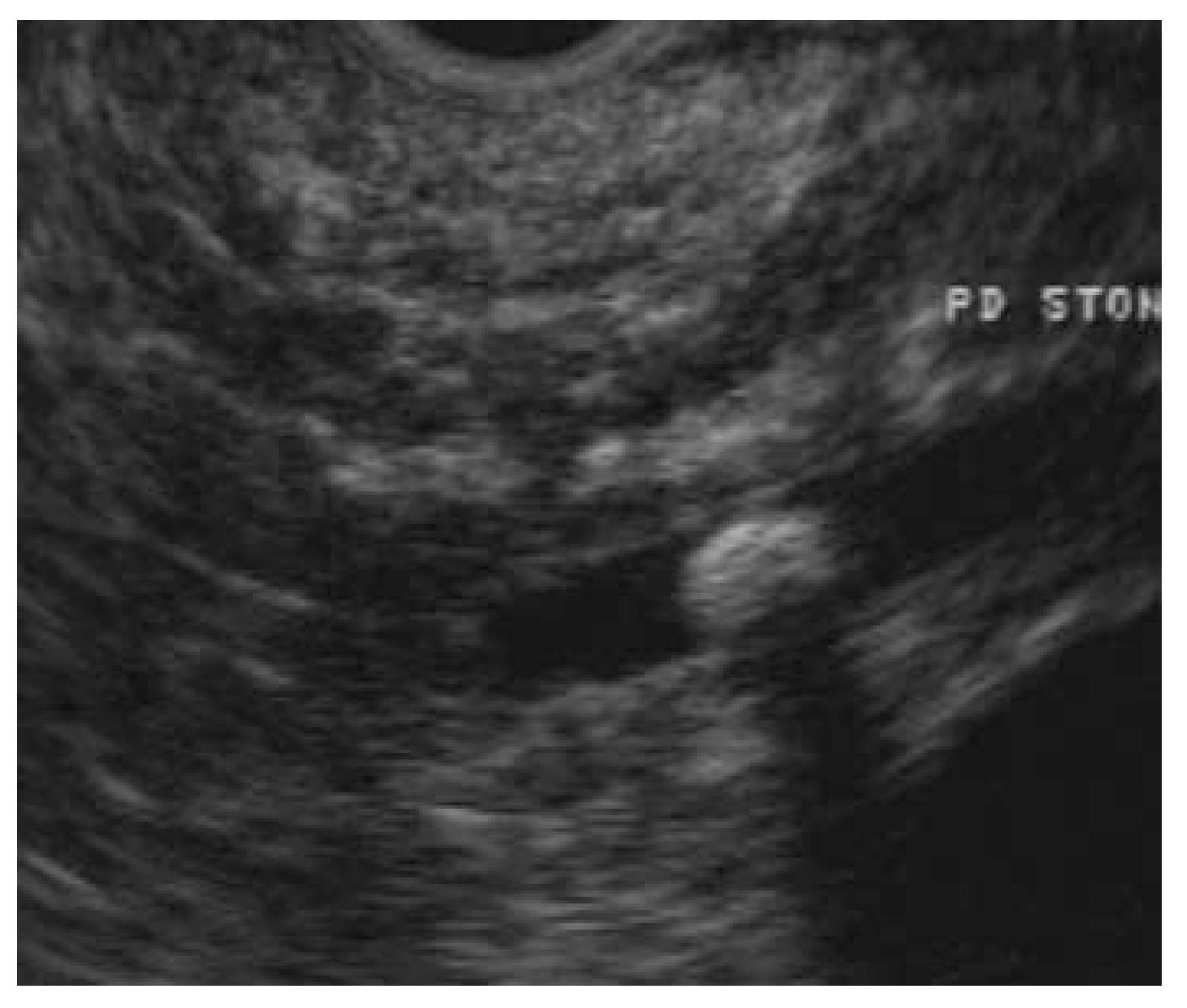
Figure 3 Linear EUS showing a shadowing stone within the pan-creatic duct (PD STONE).
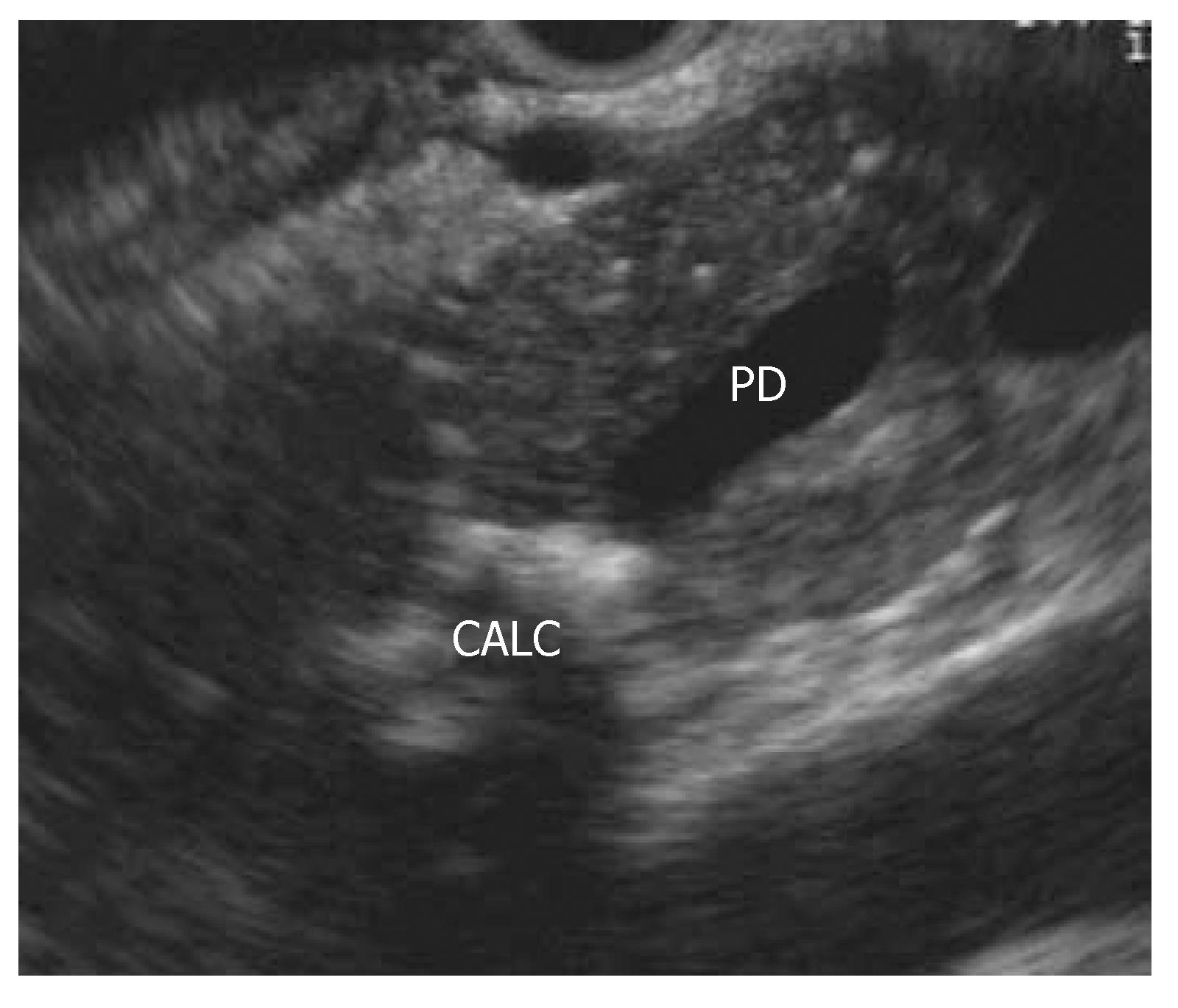
Figure 4 Linear trans-gastric EUS of the pancreas showing parench-ymal calcifications (CALC) causing dilatation of the upstream pancreatic duct (PD).
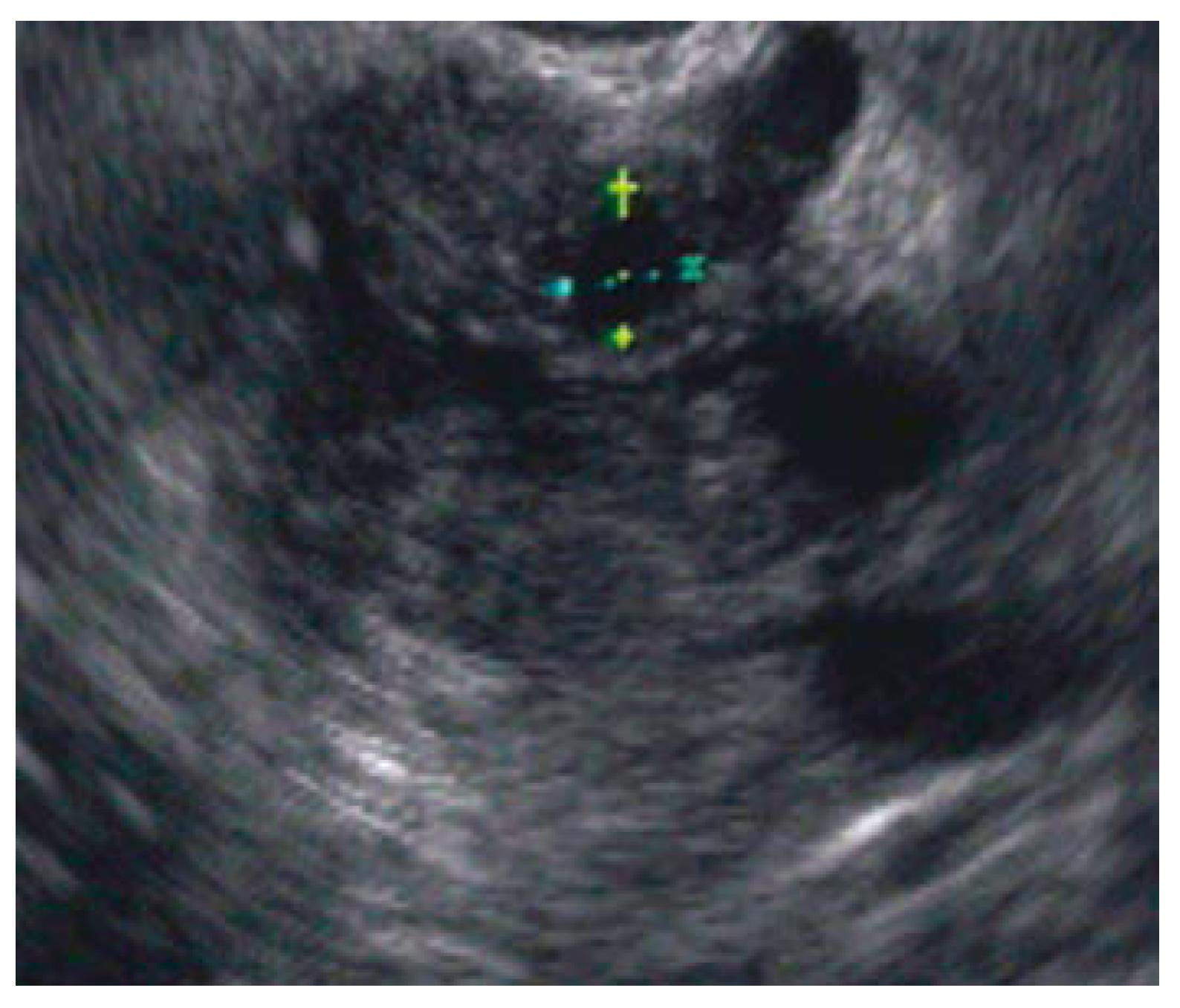
Figure 5 Linear trans-gastric EUS showing a small pancreatic cyst (labeled with mea-surement markers).
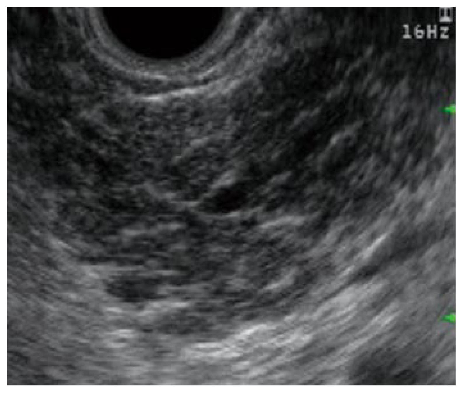
Figure 6 Linear EUS of pancreatic body with echoge-nic strands and lobularity.
- Citation: Rizk MK, Gerke H. Utility of endoscopic ultrasound in pancreatitis: A review. World J Gastroenterol 2007; 13(47): 6321-6326
- URL: https://www.wjgnet.com/1007-9327/full/v13/i47/6321.htm
- DOI: https://dx.doi.org/10.3748/wjg.v13.i47.6321


