Copyright
©2007 Baishideng Publishing Group Inc.
World J Gastroenterol. Dec 14, 2007; 13(46): 6281-6283
Published online Dec 14, 2007. doi: 10.3748/wjg.v13.i46.6281
Published online Dec 14, 2007. doi: 10.3748/wjg.v13.i46.6281
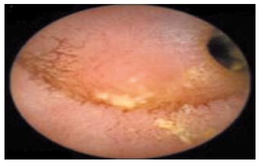
Figure 1 Image of jejeunal luminal stenosis from WCE.
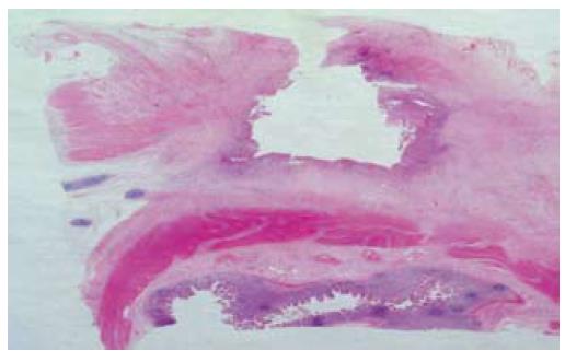
Figure 2 Photomicrograph of a deep penetrating ulcer with underlying fibrosis replacing muscularis propria, extending through the serosa adherent to an underlying small bowel loop (lower field).
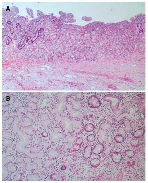
Figure 3 Photomicrographs showing (A) heterotopic gastric foveolar mucosa with a few normal small intestinal glands; and (B) scattered cells with eosinophilic cytoplasm compatible with acid-secreting cells.
- Citation: Hurley H, Cahill RA, Ryan P, Morcos AI, Redmond HP, Kiely HM. Penetrating ectopic peptic ulcer in the absence of Meckel’s diverticulum ultimately presenting as small bowel obstruction. World J Gastroenterol 2007; 13(46): 6281-6283
- URL: https://www.wjgnet.com/1007-9327/full/v13/i46/6281.htm
- DOI: https://dx.doi.org/10.3748/wjg.v13.i46.6281


