Copyright
©2007 Baishideng Publishing Group Inc.
World J Gastroenterol. Dec 14, 2007; 13(46): 6274-6276
Published online Dec 14, 2007. doi: 10.3748/wjg.v13.i46.6274
Published online Dec 14, 2007. doi: 10.3748/wjg.v13.i46.6274
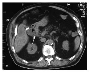
Figure 1 Abdominal CT scan six years after laparoscopic cholecystectomy.
A 4 cm encapsulated fluid collection in the gallbladder fossa (black arrow) with adjacent “cystic duct-like” collections was observed (white arrow).
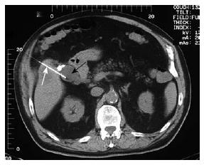
Figure 2 A CT-guided transhepatic percutaneous aspiration needle (white arrow) was seen entering the extrahepatic fluid collection (black arrow).
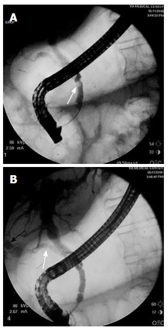
Figure 3 Calculi (white arrow) in the common bile duct were noted during the ERCP (A).
In addition, a gallbladder-like structure (black arrow) communicated with the common bile duct via the cystic duct (white arrow) was also observed (B).
- Citation: Xing J, Rochester J, Messer CK, Reiter BP, Korsten MA. A phantom gallbladder on endoscopic retrograde cholangiopancreatography. World J Gastroenterol 2007; 13(46): 6274-6276
- URL: https://www.wjgnet.com/1007-9327/full/v13/i46/6274.htm
- DOI: https://dx.doi.org/10.3748/wjg.v13.i46.6274


