Copyright
©2007 Baishideng Publishing Group Co.
World J Gastroenterol. Nov 7, 2007; 13(41): 5516-5520
Published online Nov 7, 2007. doi: 10.3748/wjg.v13.i41.5516
Published online Nov 7, 2007. doi: 10.3748/wjg.v13.i41.5516
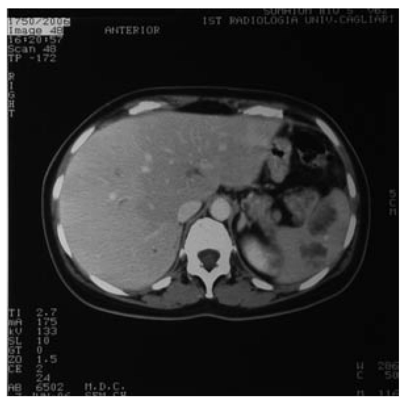
Figure 1 Two low density areas in the spleen (Axial CT-scan).
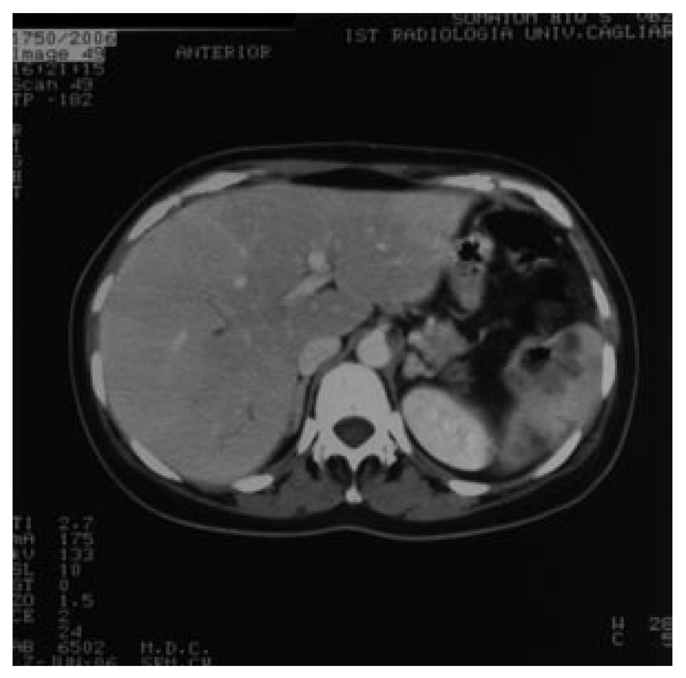
Figure 2 Wall thickening of the left flexure of the colon indistinguishable from the spleen parenchyma (Axial CT-scan).
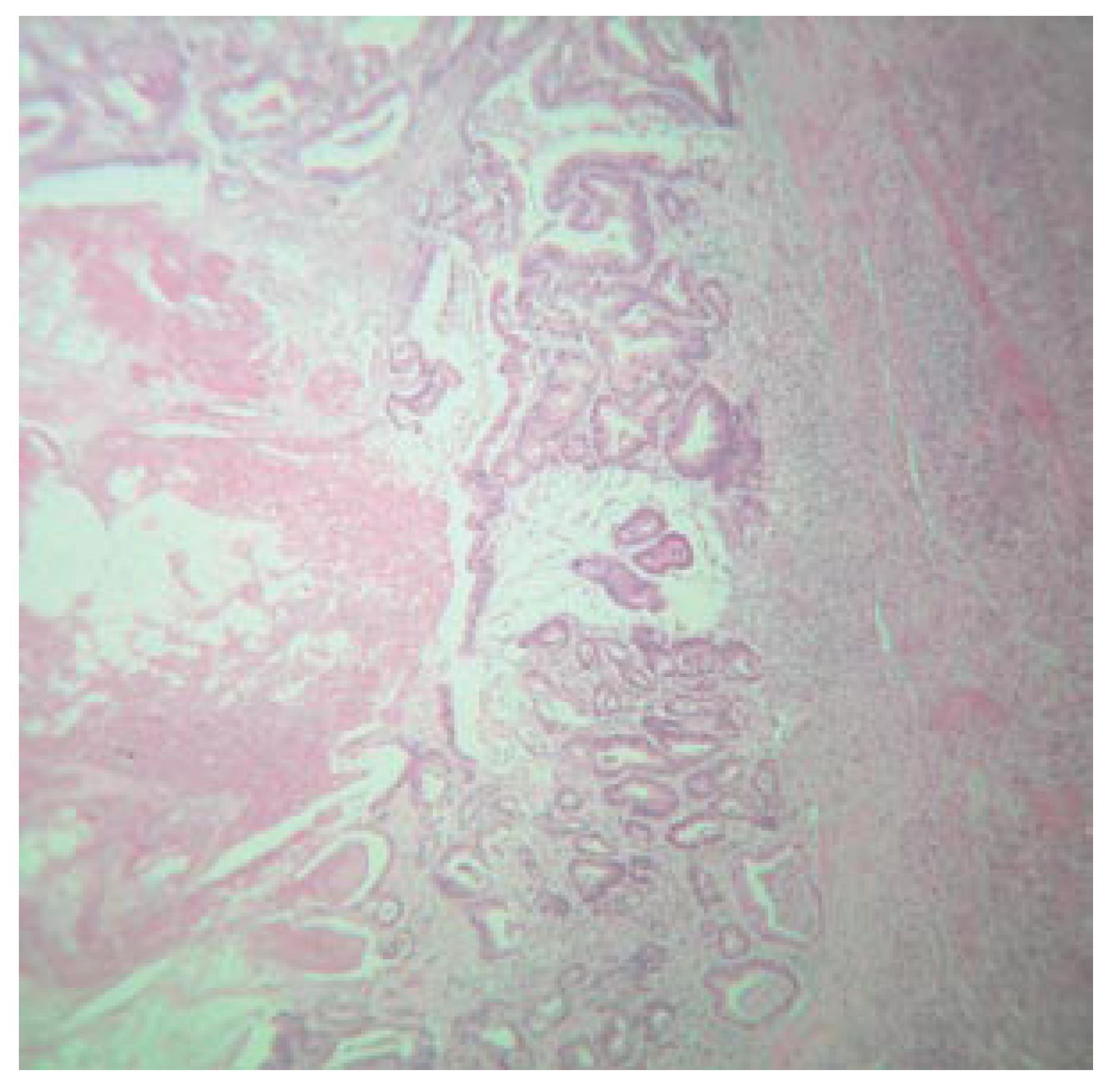
Figure 3 Splenic tumor showing glandular pattern consistent with metastasis from colonic mucinous carcinoma (HE, x 40).
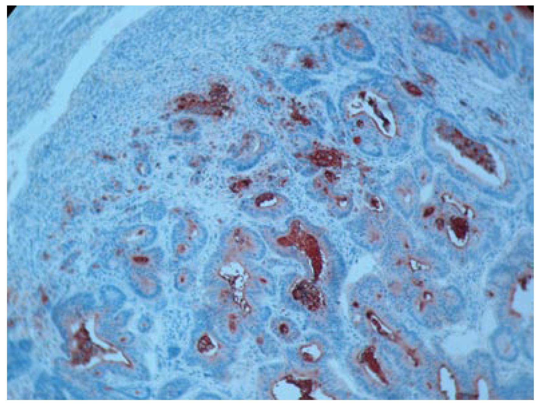
Figure 4 CEA along the luminal border of tumor cells infiltrating splenic pulp (anti-CEA monoclonal antibody staining, x 100).
- Citation: Pisanu A, Ravarino A, Nieddu R, Uccheddu A. Synchronous isolated splenic metastasis from colon carcinoma and concomitant splenic abscess: A case report and review of the literature. World J Gastroenterol 2007; 13(41): 5516-5520
- URL: https://www.wjgnet.com/1007-9327/full/v13/i41/5516.htm
- DOI: https://dx.doi.org/10.3748/wjg.v13.i41.5516


