Copyright
©2007 Baishideng Publishing Group Inc.
World J Gastroenterol. Oct 28, 2007; 13(40): 5413-5415
Published online Oct 28, 2007. doi: 10.3748/wjg.v13.i40.5413
Published online Oct 28, 2007. doi: 10.3748/wjg.v13.i40.5413
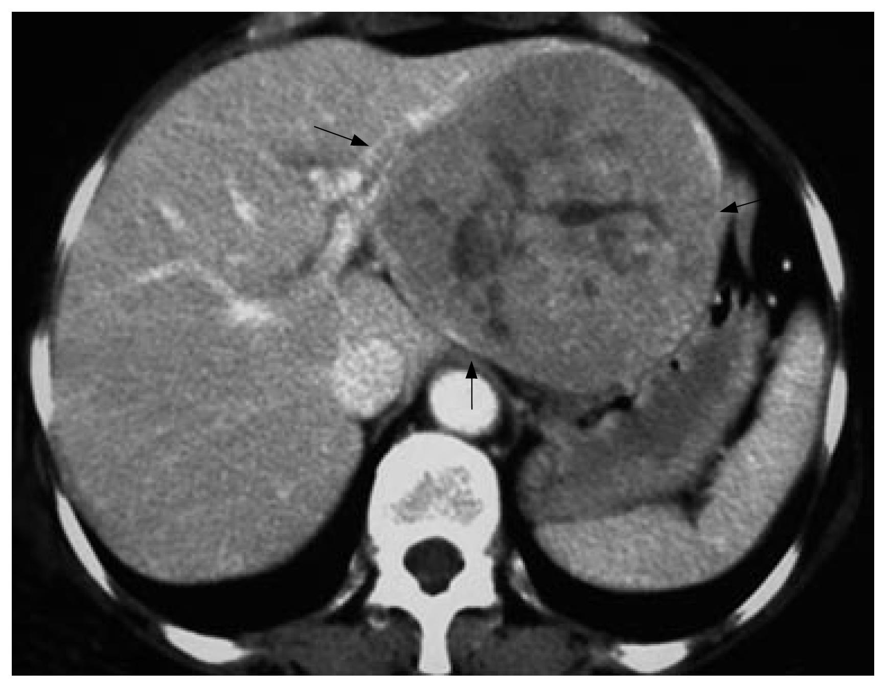
Figure 1 Contrast CT demonstrating a huge lesion at the left lobe of liver (black arrows, portal phase).
Figure 2 Whole body PET showing an isolated highly metabolic focus in the upper abdomen (white arrow) (A), non-enhanced CT (C) detecting a median density lesion in the omenta (white arrow), 18F-FDG PET (B) and fused imaging of PET/CT (D) revealing a highly metabolic lesion at the same position.
- Citation: Sun L, Guan YS, Pan WM, Chen GB, Luo ZM, Wu H. Positron emission tomography/computer tomography in guidance of extrahepatic hepatocellular carcinoma metastasis management. World J Gastroenterol 2007; 13(40): 5413-5415
- URL: https://www.wjgnet.com/1007-9327/full/v13/i40/5413.htm
- DOI: https://dx.doi.org/10.3748/wjg.v13.i40.5413


