Copyright
©2007 Baishideng Publishing Group Inc.
World J Gastroenterol. Oct 28, 2007; 13(40): 5394-5396
Published online Oct 28, 2007. doi: 10.3748/wjg.v13.i40.5394
Published online Oct 28, 2007. doi: 10.3748/wjg.v13.i40.5394
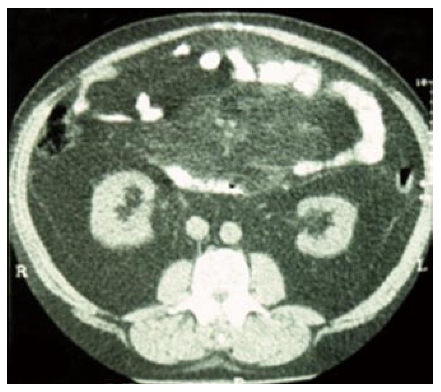
Figure 1 CT scan visualizing a mesenteric lipoma-like mass.
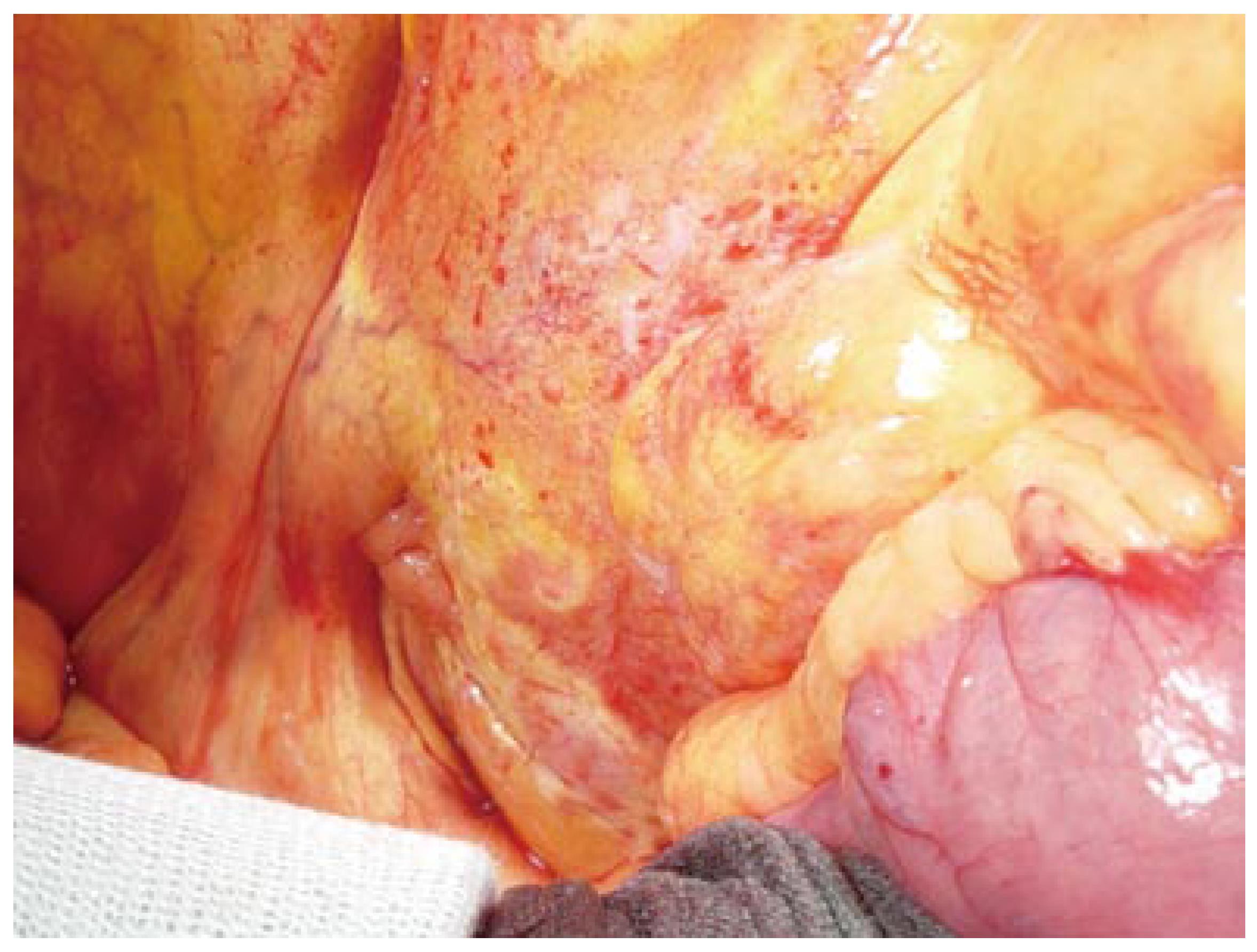
Figure 2 Intraoperative view of pseudonodular thickening of the mesentery.
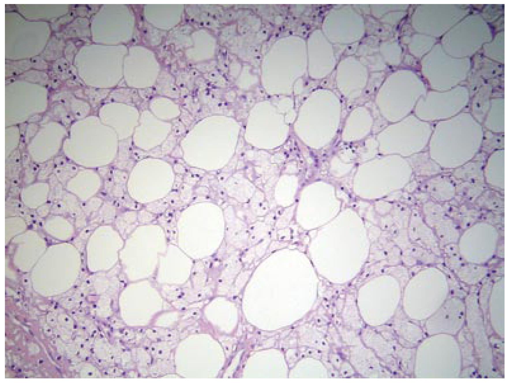
Figure 3 Mesenteric adipose tissue showing lipid necrosis and foamy macrophages (HE, x 10).
- Citation: Vettoretto N, Diana DR, Poiatti R, Matteucci A, Chioda C, Giovanetti M. Occasional finding of mesenteric lipodystrophy during laparoscopy: A difficult diagnosis. World J Gastroenterol 2007; 13(40): 5394-5396
- URL: https://www.wjgnet.com/1007-9327/full/v13/i40/5394.htm
- DOI: https://dx.doi.org/10.3748/wjg.v13.i40.5394


