Copyright
©2007 Baishideng Publishing Group Co.
World J Gastroenterol. Jan 28, 2007; 13(4): 643-646
Published online Jan 28, 2007. doi: 10.3748/wjg.v13.i4.643
Published online Jan 28, 2007. doi: 10.3748/wjg.v13.i4.643
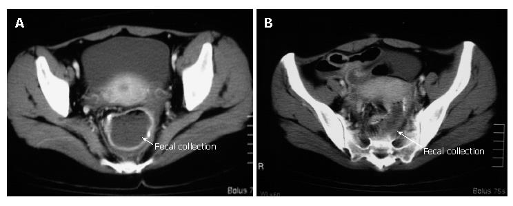
Figure 1 Abdominal computed tomography showing a massive collection of feces in the pouch (A) and proximal ileum (B).
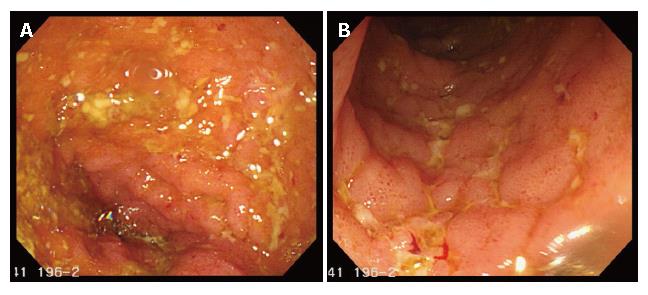
Figure 2 Pouchscopy showing inflammation conditions such as edema, granularity, friability, loss of vascular pattern, and erosions in both the pouch (A) and the pre-pouch ileum (continuous 30 cm proximal to the pouch) (B).
- Citation: Iwata T, Yamamoto T, Umegae S, Matsumoto K. Pouchitis and pre-pouch ileitis developed after restorative proctocolectomy for ulcerative colitis: A case report. World J Gastroenterol 2007; 13(4): 643-646
- URL: https://www.wjgnet.com/1007-9327/full/v13/i4/643.htm
- DOI: https://dx.doi.org/10.3748/wjg.v13.i4.643


