Copyright
©2007 Baishideng Publishing Group Co.
World J Gastroenterol. Jan 28, 2007; 13(4): 639-642
Published online Jan 28, 2007. doi: 10.3748/wjg.v13.i4.639
Published online Jan 28, 2007. doi: 10.3748/wjg.v13.i4.639
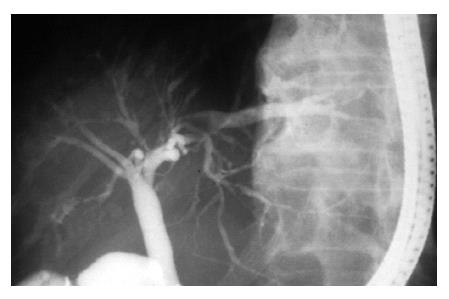
Figure 1 Endoscopic retrograde cholangiogram.
Intrahepatic biliary branches showed irregularity and extensive beading (arrows). The long arrow indicates the dilatation of the bile duct due to stenosis caused by the pressure of cysts.
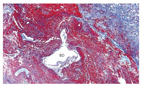
Figure 2 Extensive fibrinoid necrosis in large portal tract.
Septal bile duct is buried in the fibrinoid material (Masson’s trichrome x 1). PT: portal tract, BD: bile duct.
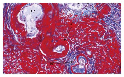
Figure 3 A smaller portal tract.
Fibrinoid material is deposited in the connective tissue, walls of the arteries and periductular connective tissue (Masson's trichrome x 20). PV: portal vein, A: artery, BD: bile duct.
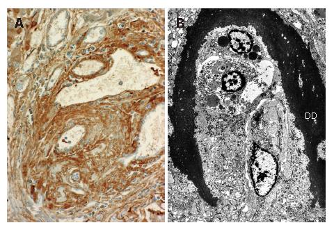
Figure 4 A: Immunohistochemical staining demonstrates the positivity of the fibrinoid material for fibrinogen; B: An electron micrograph shows dense deposit materials suggestive of fibrinoid necrosis around the small bile ducts in the portal tract of the liver (original magnification x 2000).
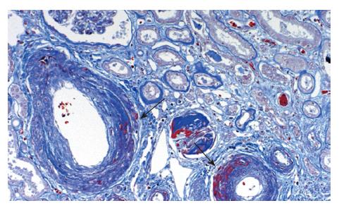
Figure 5 Kidney.
Fibrinoid necroses were seen in the walls of the interlobular arteries (arrows) (Masson’s trichrome x 10).
- Citation: Hano H, Takagi I, Nagatsuma K, Lu T, Meng C, Chiba S. An autopsy case showing massive fibrinoid necrosis of the portal tracts of the liver with cholangiographic findings similar to those of primary sclerosing cholangitis. World J Gastroenterol 2007; 13(4): 639-642
- URL: https://www.wjgnet.com/1007-9327/full/v13/i4/639.htm
- DOI: https://dx.doi.org/10.3748/wjg.v13.i4.639


