Copyright
©2007 Baishideng Publishing Group Co.
World J Gastroenterol. Jan 28, 2007; 13(4): 600-606
Published online Jan 28, 2007. doi: 10.3748/wjg.v13.i4.600
Published online Jan 28, 2007. doi: 10.3748/wjg.v13.i4.600
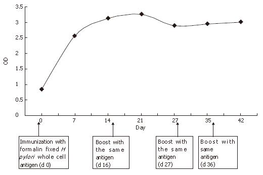
Figure 1 Kinetics of anti H pylori antibody responses in rabbits.
Female rabbits were infected with formalin-fixed H pylori whole cell antigens and blood samples were collected at various time points after H pylori infection. Sera (1:50 dilution) were subjected to ELISA for antibody titer measurement (in terms of OD). The arrows indicate the time points when rabbits were challenged with antigens (booster dose).
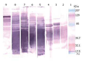
Figure 2 Western blot with whole cell extracts of H pylori against previously immunized rabbit sera and a human serum.
Lane 1: molecular weight marker; lanes 2 to 8: serum collected on d 0, 7, 14, 21, 28, 35, and 42 respectively; lane 9: positive human serum.
-
Citation: Islam K, Khalil I, Ahsan CR, Yasmin M, Nessa J. Analysis of immune responses against
H pylori in rabbits. World J Gastroenterol 2007; 13(4): 600-606 - URL: https://www.wjgnet.com/1007-9327/full/v13/i4/600.htm
- DOI: https://dx.doi.org/10.3748/wjg.v13.i4.600


