Copyright
©2007 Baishideng Publishing Group Co.
World J Gastroenterol. Oct 14, 2007; 13(38): 5154-5157
Published online Oct 14, 2007. doi: 10.3748/wjg.v13.i38.5154
Published online Oct 14, 2007. doi: 10.3748/wjg.v13.i38.5154
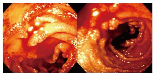
Figure 1 Endoscopy showing a vascular ectasia with active bleeding on the second portion of duodenum.
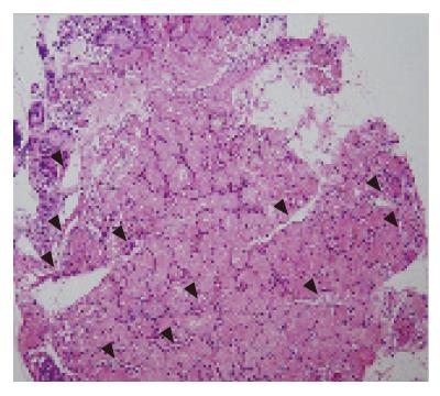
Figure 2 Biopsy from duodenal mucosa showing grossly dilated blood vessels (arrowheads) in the Brunner’s glands (HE, × 100).
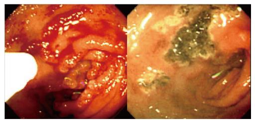
Figure 3 Induction of hemostasis by APC (Arco-2000, Soring, Germany) (left) with blood oozing controlled without complications (right).
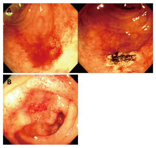
Figure 4 A one-week follow-up endoscopy showing the successfully controlled blood oozing from vascular ectasia on the second portion of the duodenum with APC (A) and a two-month follow-up endoscopy showing the ectatic vascular duodenal mucosa with no further spontaneous bleeding (B).
- Citation: Lee BJ, Park JJ, Seo YS, Kim JH, Kim A, Yeon JE, Kim JS, Byun KS, Bak YT. Upper gastrointestinal bleeding from duodenal vascular ectasia in a patient with cirrhosis. World J Gastroenterol 2007; 13(38): 5154-5157
- URL: https://www.wjgnet.com/1007-9327/full/v13/i38/5154.htm
- DOI: https://dx.doi.org/10.3748/wjg.v13.i38.5154


