Copyright
©2007 Baishideng Publishing Group Co.
World J Gastroenterol. Oct 14, 2007; 13(38): 5139-5146
Published online Oct 14, 2007. doi: 10.3748/wjg.v13.i38.5139
Published online Oct 14, 2007. doi: 10.3748/wjg.v13.i38.5139
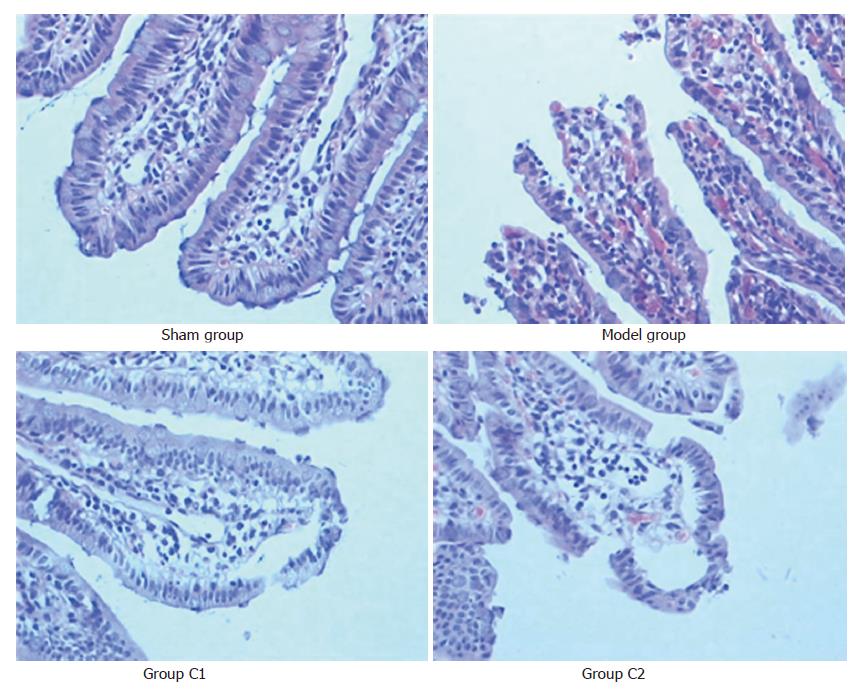
Figure 1 Microscopic appearance after hematoxylin and eosin staining (× 200).
In the model group, there are multiple erosions and bleeding in mucosal epithelial layer; the villus and glands were normal and no inflammatory cell infiltration was observed in mucosal epithelial layer in sham group; these mucosal changes are ameliorated by treatment with Cromolyn Sodium (group C1 and group C2).
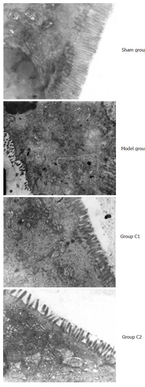
Figure 2 Changes of ultrastructure of small intestinal in each group (× 10 000).
The ultrastructure of small intestinal was normal in group S. There were seen the karyopyknosis of epithelial cell of small intestine in group M, and the nuclear membrane was more irregularity, and the swelling microvillus became shorter and thicker, most of the microvillus were shedding. The nucleus of epithelial cell of small intestine in group C1 and C2 was deflated, the nuclear membrane was irregularity, and the microvillus became shorter and light swelling.
Figure 3 Ultrastructure of intestinal mucosal mast cells of rats in each group.
There are abundant vacuolus with a reduction of granulation in their endochylema in model group; there are filled with granulation endochylema and there is no vacuolus in their endochylema in sham group, these changes of ultrastructure are ameliorated by treatment with Cromolyn Sodium (group C1 and C2).
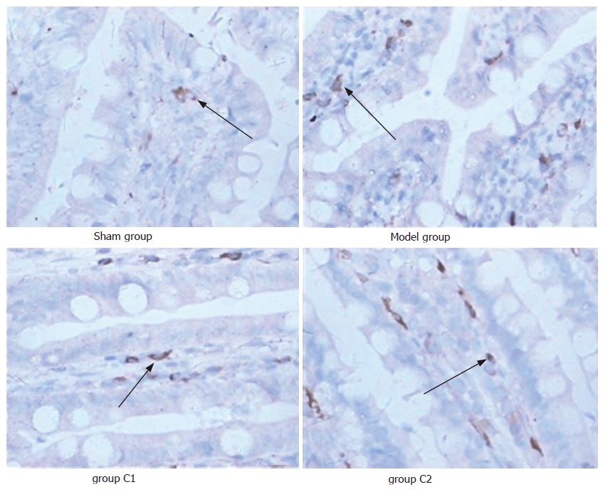
Figure 4 Immunohistochemical detection of tryptase in small intestinal of rats in each group (×400).
Expression of tryptase and number of IMMC are increased in the model group; these changes were ameliorated by treatment with Cromolyn Sodium (group C1 and C2).
- Citation: Hei ZQ, Gan XL, Luo GJ, Li SR, Cai J. Pretreatment of cromolyn sodium prior to reperfusion attenuates early reperfusion injury after the small intestine ischemia in rats. World J Gastroenterol 2007; 13(38): 5139-5146
- URL: https://www.wjgnet.com/1007-9327/full/v13/i38/5139.htm
- DOI: https://dx.doi.org/10.3748/wjg.v13.i38.5139


