Copyright
©2007 Baishideng Publishing Group Co.
World J Gastroenterol. Oct 14, 2007; 13(38): 5127-5132
Published online Oct 14, 2007. doi: 10.3748/wjg.v13.i38.5127
Published online Oct 14, 2007. doi: 10.3748/wjg.v13.i38.5127
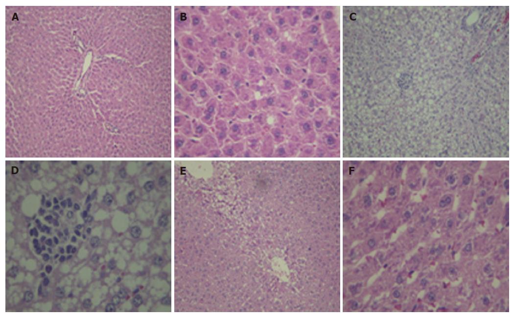
Figure 1 Hematoxylin and eosin staining of liver tissue.
A, B: control; C, D: NASH, fed with 100% fat diet group showed macrovesicular steatosis, ballooning changes, Mallory bodies, hepatocyte necrosis, and infiltration of inflammatory cells; E, F: NASH + NAC20, showed the improvement in steatosis and necroinflammation (A, C, E: × 10; B, D, F: × 40 ).
- Citation: Thong-Ngam D, Samuhasaneeto S, Kulaputana O, Klaikeaw N. N-acetylcysteine attenuates oxidative stress and liver pathology in rats with non-alcoholic steatohepatitis. World J Gastroenterol 2007; 13(38): 5127-5132
- URL: https://www.wjgnet.com/1007-9327/full/v13/i38/5127.htm
- DOI: https://dx.doi.org/10.3748/wjg.v13.i38.5127


