Copyright
©2007 Baishideng Publishing Group Co.
World J Gastroenterol. Oct 7, 2007; 13(37): 5035-5037
Published online Oct 7, 2007. doi: 10.3748/wjg.v13.i37.5035
Published online Oct 7, 2007. doi: 10.3748/wjg.v13.i37.5035
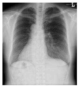
Figure 1 A chest radiograph showing dextrocardia and a right subphrenic gastric bubble.
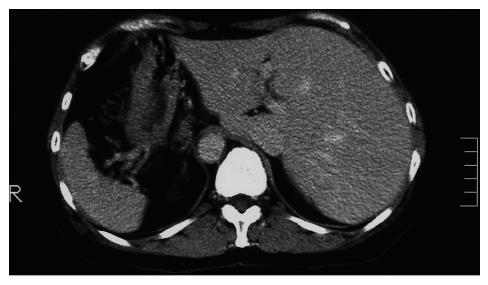
Figure 2 Computed tomography disclosing complete transposition of abdominal viscera.
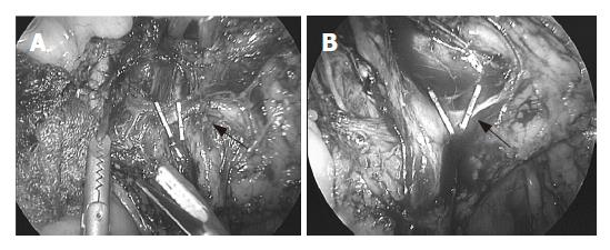
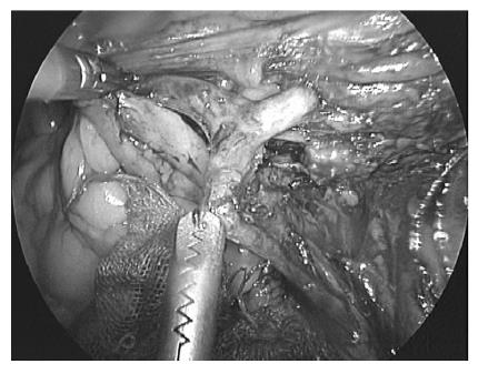
Figure 3 Initial laparoscopic vascular procedures identifying the ileocolic artery and dividing it at its root (arrow) (A) and ileocolic vein after exposure of the superior mesenteric vein (arrow) (B).
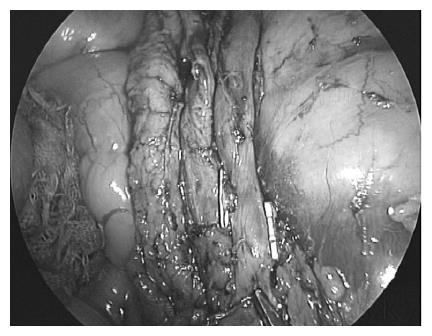
Figure 4 Radical lymphadenectomy along the superior mesenteric artery.
Figure 5 Division of the left branch of the middle colic artery.
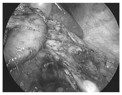
Figure 6 Full mobilization of the ascending colon including the tumor.
- Citation: Fujiwara Y, Fukunaga Y, Higashino M, Tanimura S, Takemura M, Tanaka Y, Osugi H. Laparoscopic hemicolectomy in a patient with situs inversus totalis. World J Gastroenterol 2007; 13(37): 5035-5037
- URL: https://www.wjgnet.com/1007-9327/full/v13/i37/5035.htm
- DOI: https://dx.doi.org/10.3748/wjg.v13.i37.5035


