Copyright
©2007 Baishideng Publishing Group Co.
World J Gastroenterol. Sep 7, 2007; 13(33): 4514-4516
Published online Sep 7, 2007. doi: 10.3748/wjg.v13.i33.4514
Published online Sep 7, 2007. doi: 10.3748/wjg.v13.i33.4514
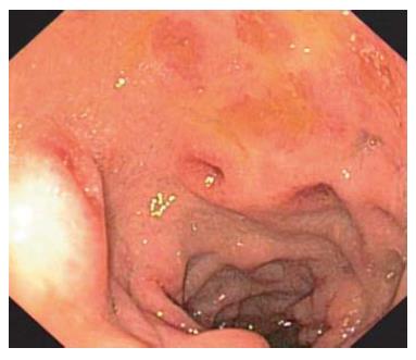
Figure 1 Inflammation of the duodenal mucosa with ulcerous lesions can be seen.
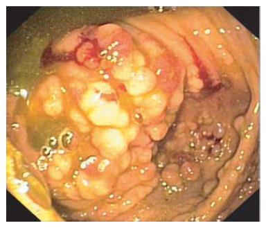
Figure 2 Diffuse polypoid lesions are present in the colon.
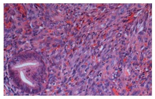
Figure 3 Proliferation of spindle cells is seen in the lamina propria.
Slit-like spaces are present, with red blood cells between them.
- Citation: Zoufaly A, Schmiedel S, Lohse A, van Lunzen J. Intestinal Kaposi’s sarcoma may mimic gastrointestinal stromal tumor in HIV infection. World J Gastroenterol 2007; 13(33): 4514-4516
- URL: https://www.wjgnet.com/1007-9327/full/v13/i33/4514.htm
- DOI: https://dx.doi.org/10.3748/wjg.v13.i33.4514


