Copyright
©2007 Baishideng Publishing Group Co.
World J Gastroenterol. Aug 21, 2007; 13(31): 4185-4191
Published online Aug 21, 2007. doi: 10.3748/wjg.v13.i31.4185
Published online Aug 21, 2007. doi: 10.3748/wjg.v13.i31.4185
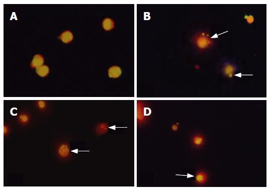
Figure 1 MKN45 cells, treated or not treated with FA-2-b-β and AZT.
A: Untreated cells; B-D: FA-2-b-β (5 mg/L) + AZT 5, 10 and 20 mg/L for 72 h exposure, apoptotic body (B), nuclear fragmentation (C) and chromatin condensation (D) (Acridine orange staining, × 100).
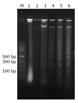
Figure 2 Agarose gel electrophoresis of DNA of MKN45 cells.
M: marker, Lane 1: control; Lane 2-5: MKN45 cells treated by FA-2-b-β (5 mg/L), AZT (2.5 mg/L), FA-2-b-β (5 mg/L) + AZT 5, 10 and 20 mg/L for 72 h, respectively.
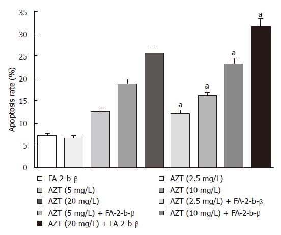
Figure 3 Effect of FA-2-b-β (5 mg/L) and AZT on apoptosis rate in MKN45 cell for 72 h (aP < 0.
05).
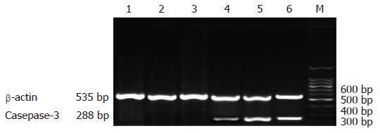
Figure 4 Expression of Caspase-3 mRNA in MKN45 cells treated with AZT and FA-2-b-β.
M: marker; Lane 1: control; Lane 2-6: MKN45 cells treated by FA-2-b-β (5 mg/L), AZT (2.5 mg/L), FA-2-b-β (5 mg/L) + AZT 5, 10, 20 mg/L respectively for 72 h.
Figure 5 Expression of Bcl-2 mRNA in MKN45 cells treated with AZT and FA-2-b-β.
M: marker; Lane 1-5: MKN45 cells treated by FA-2-b-β (5 mg/L) + AZT 20, 10 and 5 mg/L, AZT (2.5 mg/L), FA-2-b-β (5 mg/L), respectively for 72 h; Lane 6: control.
- Citation: Sun YQ, Guo TK, Xi YM, Chen C, Wang J, Wang ZR. Effects of AZT and RNA-protein complex (FA-2-b-β) extracted from Liang Jin mushroom on apoptosis of gastric cancer cells. World J Gastroenterol 2007; 13(31): 4185-4191
- URL: https://www.wjgnet.com/1007-9327/full/v13/i31/4185.htm
- DOI: https://dx.doi.org/10.3748/wjg.v13.i31.4185


