Copyright
©2007 Baishideng Publishing Group Co.
World J Gastroenterol. Aug 14, 2007; 13(30): 4154-4155
Published online Aug 14, 2007. doi: 10.3748/wjg.v13.i30.4154
Published online Aug 14, 2007. doi: 10.3748/wjg.v13.i30.4154
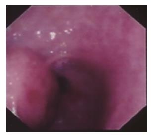
Figure 1 Upper GI endoscopy showing a mucosal lesion in the gastric antrum being suspected for lipomatous tumor.
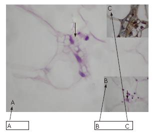
Figure 2 Multivacuolated (A) and univacuolated (B) lipoblasts as well as a multivacuolated lipoblast positive immunohistostain for S-100 protein (C).
- Citation: Tepetes K, Christodoulidis G, Spyridakis ME, Nakou M, Koukoulis G, Hatzitheofilou K. Liposarcoma of the stomach: A rare case report. World J Gastroenterol 2007; 13(30): 4154-4155
- URL: https://www.wjgnet.com/1007-9327/full/v13/i30/4154.htm
- DOI: https://dx.doi.org/10.3748/wjg.v13.i30.4154


