Copyright
©2007 Baishideng Publishing Group Co.
World J Gastroenterol. Aug 14, 2007; 13(30): 4147-4148
Published online Aug 14, 2007. doi: 10.3748/wjg.v13.i30.4147
Published online Aug 14, 2007. doi: 10.3748/wjg.v13.i30.4147
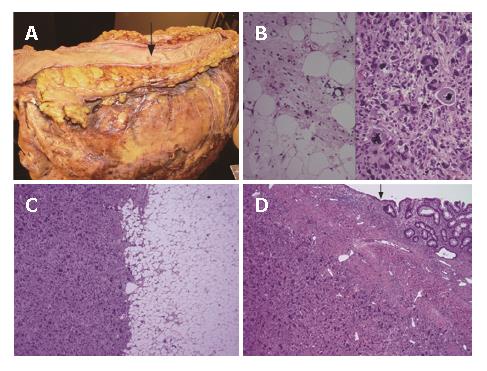
Figure 1 A: Gross picture showing a 25 cm dedifferentiated liposarcoma arising from the sigmoid mesocolon.
Notice the bowel mucosa is unremarkable except for a small ulcer (arrow); B: The well-differentiated component showing numerous lipoblasts and fibrous septae. The dedifferentiated component resembles a malignant fibrous histiocytoma with pleomorphic nuclei and tumor giant cells (HE, x 400); C: The area showing an abrupt transition from the well-differentiated component to the dedifferentiated component (HE, x 40); D: The dedifferentiated component invading into the bowel wall. The arrow points to the overlying bowel mucosa (HE, x 40).
- Citation: Winn B, Gao J, Akbari H, Bhattacharya B. Dedifferentiated liposarcoma arising from the sigmoid mesocolon: A case report. World J Gastroenterol 2007; 13(30): 4147-4148
- URL: https://www.wjgnet.com/1007-9327/full/v13/i30/4147.htm
- DOI: https://dx.doi.org/10.3748/wjg.v13.i30.4147


