Copyright
©2007 Baishideng Publishing Group Co.
World J Gastroenterol. Aug 14, 2007; 13(30): 4035-4041
Published online Aug 14, 2007. doi: 10.3748/wjg.v13.i30.4035
Published online Aug 14, 2007. doi: 10.3748/wjg.v13.i30.4035
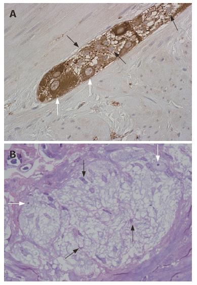
Figure 1 A: Human colonic myenteric plexus showing that neurons (white arrows) are less numerous with respect to EGC (black arrows) (NSE immunostaining, x 40); B: Semithin section of human colonic submucosal plexus, showing the preponderance of EGC (black arrows) with respect to the enteric neurons (white arrows) (Toluidine blue, x 40).
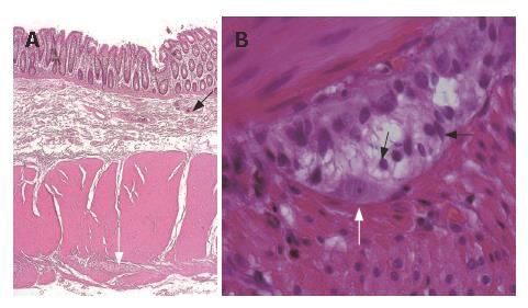
Figure 2 A: Full thickness section of the human colon, showing the submucosal (black arrow) and the myenteric plexus (white arrow) (HE, x10); B: Human myenteric ganglion, showing numerous EGC (black arrows) and an enteric neuron (white arrow) (HE, x 100).
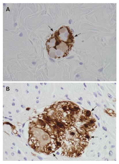
Figure 3 EGC (arrows) tightly packed around enteric neurons in a submucosal (A) and a myenteric ganglion (B) (S100 immunostaining, x 40).
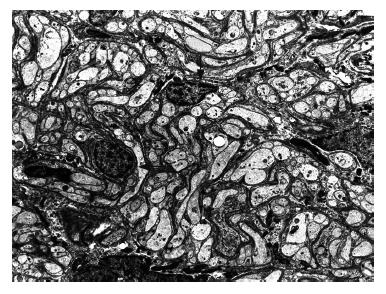
Figure 4 Electron microscopic image of glial cells (arrows) in a human colonic myenteric ganglion (x 1900).
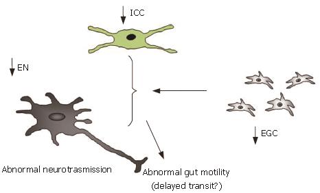
Figure 5 Putative mechanisms linked to the decrease of enteric glial cells (EGC), leading to abnormal gut motility.
EN: enteric neurons; ICC: interstitial cells of Cajal.
- Citation: Bassotti G, Villanacci V, Fisogni S, Rossi E, Baronio P, Clerici C, Maurer CA, Cathomas G, Antonelli E. Enteric glial cells and their role in gastrointestinal motor abnormalities: Introducing the neuro-gliopathies. World J Gastroenterol 2007; 13(30): 4035-4041
- URL: https://www.wjgnet.com/1007-9327/full/v13/i30/4035.htm
- DOI: https://dx.doi.org/10.3748/wjg.v13.i30.4035


